IF du Neurocentre
IF1234567891011121314151617181920253035404550
Nombre00383110240001111102120303
%007187330590003333305350707

45 publications


07/2025 | Mol Psychiatry
Nutritional interventions to counteract the detrimental consequences of early-life stress.
Geertsema J, Juncker HG, Wilmes L, Burchell GL, de Rooij SR, van Goudoever JB, O'Riordan KJ, Clarke G, Cryan JF, Korosi A
doi: 10.1038/s41380-025-03020-1

Abstract:
Exposure to stress during sensitive developmental periods comes with long term consequences for neurobehavioral outcomes and increases vulnerability to psychopathology later in life. While we have advanced our understanding of the mechanisms underlying the programming effects of early-life stress (ES), these are not yet fully understood and often hard to target, making the development of effective interventions challenging. In recent years, we and others have suggested that nutrition might be instrumental in modulating and possibly combatting the ES-induced increased risk to psychopathologies and neurobehavioral impairments. Nutritional strategies are very promising as they might be relatively safe, cheap and easy to implement. Here, we set out to comprehensively review the existing literature on nutritional interventions aimed at counteracting the effects of ES on neurobehavioral outcomes in preclinical and clinical settings. We identified eighty six rodent and ten human studies investigating a nutritional intervention to ameliorate ES-induced impairments. The human evidence to date, is too few and heterogeneous in terms of interventions, thus not allowing hard conclusions, however the preclinical studies, despite their heterogeneity in terms of designs, interventions used, and outcomes measured, showed nutritional interventions to be promising in combatting ES-induced impairments. Furthermore, we discuss the possible mechanisms involved in the beneficial effects of nutrition on the brain after ES, including neuroinflammation, oxidative stress, hypothalamus-pituitary-adrenal axis regulation and the microbiome-gut-brain axis. Lastly, we highlight the critical gaps in our current knowledge and make recommendations for future research to move the field forward.


02/11/2024 | Cell Metab
Gut microbiota regulates stress responsivity via the circadian system.
Tofani GSS, Leigh SJ, Gheorghe CE, Bastiaanssen TFS, Wilmes L, Sen P, Clarke G, Cryan JF

Abstract:
Stress and circadian systems are interconnected through the hypothalamic-pituitary-adrenal (HPA) axis to maintain responses to external stimuli. Yet, the mechanisms of how such signals are orchestrated remain unknown. Here, we uncover the gut microbiota as a regulator of HPA-axis rhythmicity. Microbial depletion disturbs the brain transcriptome and metabolome in stress-responding pathways in the hippocampus and amygdala across the day. This is coupled with a dysregulation of the circadian pacemaker in the brain that results in perturbed glucocorticoid rhythmicity. The resulting hyper-activation of the HPA axis at the sleep/wake transition drives time-of-day-specific impairments of the stress response and stress-sensitive behaviors. Finally, microbiota transplantation confirmed that diurnal oscillations of gut microbes underlie altered glucocorticoid secretion and that L. reuteri is a candidate strain for such effects. Our data offer compelling evidence that the microbiota regulates stress responsiveness in a circadian manner and is necessary to respond adaptively to stressors throughout the day.


11/2024 | neurobiol stress
Sex specific gut-microbiota signatures of resilient and comorbid gut-brain phenotypes induced by early life stress.
Wilmes L, Caputi V, Bastiaanssen TFS, Collins JM, Crispie F, Cotter PD, Dinan TG, Cryan JF, Clarke G, O'Mahony SM

Abstract:
BACKGROUND: Alterations in gut-brain axis communication pathways and the gut microbiota ecosystem caused by early life stress have been extensively described as critical players in the pathophysiology of stress-induced disorders. However, the extent to which stress-induced gut microbiota alterations manifest in early life and contribute to the sex-specific susceptibility to distinct gut-brain phenotypes in adulthood has yet to be defined. METHODS: Male and female Sprague-Dawley rat offspring underwent maternal separation (3h/day from postnatal day 2-12). Faecal samples were collected before weaning for gut microbiota 16S rRNA sequencing and metabolomic analysis. Visceral pain sensitivity and negative valence behaviours were assessed in adulthood using colorectal distension and the forced swim test respectively. Behavioural data were processed in a two-step cluster analysis to identify groupings within the dataset. Multi-omics analysis was carried out to investigate if the microbial signatures following early life stress were already defined according to the membership of the adult behavioural phenotypes. RESULTS: Maternal separation resulted in increased visceral hypersensitivity while showing a trend for a sex-dependent increase in negative valence behaviour in adulthood. The cluster analysis revealed four clusters within the dataset representing distinct pathophysiological domains reminiscent of the behavioural consequences of early-life stress: 1. resilient, 2. pain, 3. immobile and 4. comorbid. The early life gut microbiota of each of these clusters show distinct alterations in terms of diversity, genus level differential abundance, and functional modules. Multi-omic integrations points towards a role for different metabolic pathways underlying each cluster-specific phenotype. CONCLUSION: Our study is the first to identify distinct phenotypes defined by susceptibility or resilience to gut-brain dysfunction induced by early life stress. The gut microbiota in early life shows sex-dependent alterations in each cluster that precede specific behavioural phenotypes in adulthood. Future research is warranted to determine the causal relationship between early-life stress-induced changes in the gut microbiota and to understand the trajectory leading to the manifestation of different behavioural phenotypes in adulthood.


10/2024 | BioRxiv
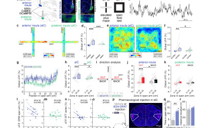
Dopamine transmission in the anterior insula shapes the neural coding of anxiety
Couderc Y, Dhanireddy T, Vardiero G, Garg A, Ricci D, D'almeida M, Nicolas C, Habchi T, Wu KY, Gjorgjieva J, Li Y, Valjent E, Beyeler A
doi: https://doi.org/10.1101/2024.10.25.620186

Abstract:
The insular cortex (or insula), and particularly its anterior region, plays a crucial role in the control of emotional valence and anxiety (Etkin & Wager, 2007; Méndez-Ruette et al., 2019; Nicolas et al., 2023). While dopamine neurotransmission is known to modulate anxiety levels in humans (Hjorth et al., 2021) and animal models (de la Mora et al., 2010; Bananej et al., 2012; Zarrindast & Khakpai, 2015; DeGroot et al., 2020; Godino et al., 2023), its regulatory effects on the anterior insula remained unexplored. Here, using a multifaceted approach, we uncovered how dopamine shapes anterior insula function in anxiety and valence processing. First, we revealed a high density of neurons expressing type-1 dopamine receptors (D1) in the insula, particularly important in the anterior insula, and seven times greater than the density of neurons expressing type-2 dopamine receptors (D2). Few neurons co-expressed Drd1 and Drd2 mRNAs in the anterior and posterior insula, and the density of Drd1+ neurons in the anterior insula was twice higher among inhibitory neurons than excitatory neurons. Second, we found that pharmacological activation of D1 in the anterior insula is anxiogenic, suggesting a direct link between insular dopamine signaling and anxiety-related behaviors. Using fiber-photometry recordings, we identified that the amplitude of dopamine release onto D1+ neurons in the anterior insula while mice were in anxiogenic spaces or receiving mild foot shocks was both positively correlated with mice level of trait anxiety. Population dynamics and deep-learning analyses of anterior insula single-unit recordings uncovered distinct coding patterns of anxiety-provoking and safe environments, as well as tastants of positive and negative valence. Remarkably, systemic D1 activation, which heightens anxiety- related behaviors, dampens this coding dichotomy by increasing coding variability for protected spaces while increasing the coding reliability for anxiogenic spaces. Interestingly, the coding reliability of anxiogenic areas was positively correlated with mice level of trait anxiety, and we observed a trend towards a positive correlation between the coding reliability of a negative tastants, and mice level of anxiety. Altogether, our findings provide a new model of neural population coding of anxiety and emotional valence and unravel D1-dependent coding mechanisms in the mouse anterior insula.




25/09/2024 | Int J Biochem Cell Biol
Classical psychedelics' action on brain monoaminergic systems.
Butler JJ, Ricci D, Aman C, Beyeler A, De Deurwaerdere P

Abstract:
The study of the mechanism of action of classical psychedelics has gained significant interest due to their clinical potential in the treatment of several psychiatric conditions, including major depressive and anxiety disorders. These drugs bind 5-hydroxytryptamine receptors (5-HTR) including 5-HT(1A)R, 5-HT(2A)R, 5-HT(2B)R, and/or 5-HT(2)(C)R, as well as other targets. 5-HTRs regulate the activity of ascending monoaminergic neurons, a mechanism primarily involved in the action of classical antidepressant drugs, antipsychotics, and drugs of abuse. Sparse neurochemical data have been produced on the control of monoaminergic neuron activity in response to classical psychedelics. Here we review the available data in order to determine whether classical psychedelics have specific neurochemical effects on serotonergic, dopaminergic, and noradrenergic neurons. The data show that these drugs have disparate effects on each monoaminergic system, demonstrating a complex response with state-dependent and region-specific effects. For instance, several psychedelics inhibit the firing of serotonergic neurons, although this is not necessarily associated with a decrease in serotonin release in all regions. Noradrenergic neuron spontaneous activity also appears to be inhibited by psychedelics, also not necessarily associated with a decrease in noradrenaline release in all regions. Psychedelics influence on dopaminergic systems is also complex as the above-mentioned 5-HTRs may have opposing effects on dopaminergic neuron activity, in a state-dependent manner. There is an apparent lack of clear neuronal signature induced by psychedelics on monoaminergic neuron activity despite specific recurrent mechanisms. This review provides a current summary of the action of psychedelics on monoamine neuromodulators serotonin, dopamine and noradrenaline, compiling reoccurring and contradictory findings demonstrating that a monoamine signature of psychedelics, if applicable, would be state- and region-dependant.


Abstract:
Single administration of low-dose ketamine has both acute and sustained anti-depressant effects. Sustained effect is associated with restoration of glutamatergic synapses in medial prefrontal cortic (mFPC) neurons. Ketamine induced profound changes in a number of molecular pathways in a mouse model for chronic stress. Cell-cell communication analyses predicted that planar-cell-polarity (PCP) signaling was decreased after chronic administration of corticosterone but increased following ketamine administration in most of the excitatory neurons. Similar decrease of PCP signaling in excitatory neurons was predicted in dorsolateral prefrontal cortical (dl-PFC) neurons of patients with major depressive disorder (MDD). We showed that the basolateral amygdala (BLA)-projecting infralimbic prefrontal cortex (IL PFC) neurons regulate immobility time in the tail suspension test and food consumption. Conditionally knocking out Celsr2 and Celsr3 or Prickle2 in the BLA-projecting IL PFC neurons abolished ketamine-induced synapse restoration and behavioral remission. Therefore, PCP proteins in IL PFC-BLA neurons mediate synapse restoration induced by of low-dose ketamine.


15/04/2024 | Curr Biol
Cannabinoids regulate an insula circuit controlling water intake.
Zhao Z, Covelo A, Couderc Y, Mitra A, Varilh M, Wu Y, Jacky D, Fayad R, Cannich A, Bellocchio L, Marsicano G, Beyeler A
doi: 10.1016/j.cub.2024.03.053

Abstract:
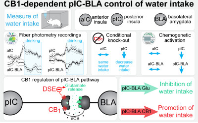
The insular cortex, or insula, is a large brain region involved in the detection of thirst and the regulation of water intake. However, our understanding of the topographical, circuit, and molecular mechanisms for controlling water intake within the insula remains parcellated. We found that type-1 cannabinoid (CB(1)) receptors in the insular cortex cells participate in the regulation of water intake and deconstructed the circuit mechanisms of this control. Topographically, we revealed that the activity of excitatory neurons in both the anterior insula (aIC) and posterior insula (pIC) increases in response to water intake, yet only the specific removal of CB(1) receptors in the pIC decreases water intake. Interestingly, we found that CB(1) receptors are highly expressed in insula projections to the basolateral amygdala (BLA), while undetectable in the neighboring central part of the amygdala. Thus, we recorded the neurons of the aIC or pIC targeting the BLA (aIC-BLA and pIC-BLA) and found that they decreased their activity upon water drinking. Additionally, chemogenetic activation of pIC-BLA projection neurons decreased water intake. Finally, we uncovered CB(1)-dependent short-term synaptic plasticity (depolarization-induced suppression of excitation [DSE]) selectively in pIC-BLA, compared with aIC-BLA synapses. Altogether, our results support a model where CB(1) receptor signaling promotes water intake by inhibiting the pIC-BLA pathway, thereby contributing to the fine top-down control of thirst responses.


10/2023 | J Physiol
The impact of acute and chronic stress on gastrointestinal physiology and function: a microbiota-gut-brain axis perspective.
Leigh SJ, Uhlig F, Wilmes L, Sanchez-Diaz P, Gheorghe CE, Goodson MS, Kelley-Loughnane N, Hyland NP, Cryan JF, Clarke G
doi: 10.1113/JP281951

Abstract:
The physiological consequences of stress often manifest in the gastrointestinal tract. Traumatic or chronic stress is associated with widespread maladaptive changes throughout the gut, although comparatively little is known about the effects of acute stress. Furthermore, these stress-induced changes in the gut may increase susceptibility to gastrointestinal disorders and infection, and impact critical features of the neural and behavioural consequences of the stress response by impairing gut-brain axis communication. Understanding the mechanisms behind changes in enteric nervous system circuitry, visceral sensitivity, gut barrier function, permeability, and the gut microbiota following stress is an important research objective with pathophysiological implications in both neurogastroenterology and psychiatry. Moreover, the gut microbiota has emerged as a key aspect of physiology sensitive to the effects of stress. In this review, we focus on different aspects of the gastrointestinal tract including gut barrier function as well as the immune, humoral and neuronal elements involved in gut-brain communication. Furthermore, we discuss the evidence for a role of stress in gastrointestinal disorders. Existing gaps in the current literature are highlighted, and possible avenues for future research with an integrated physiological perspective have been suggested. A more complete understanding of the spatial and temporal dynamics of the integrated host and microbial response to different kinds of stressors in the gastrointestinal tract will enable full exploitation of the diagnostic and therapeutic potential in the fast-evolving field of host-microbiome interactions.


21/08/2023 | Nat Commun
Linking emotional valence and anxiety in a mouse insula-amygdala circuit.
Nicolas C, Ju A, Wu Y, Eldirdiri H, Delcasso S, Couderc Y, Fornari C, Mitra A, Supiot L, Verite A, Masson M, Rodriguez-Rozada S, Jacky D, Wiegert JS, Beyeler A

Abstract:
Responses of the insular cortex (IC) and amygdala to stimuli of positive and negative valence are altered in patients with anxiety disorders. However, neural coding of both anxiety and valence by IC neurons remains unknown. Using fiber photometry recordings in mice, we uncover a selective increase of activity in IC projection neurons of the anterior (aIC), but not posterior (pIC) section, when animals are exploring anxiogenic spaces, and this activity is proportional to the level of anxiety of mice. Neurons in aIC also respond to stimuli of positive and negative valence, and the strength of response to strong negative stimuli is proportional to mice levels of anxiety. Using ex vivo electrophysiology, we characterized the IC connection to the basolateral amygdala (BLA), and employed projection-specific optogenetics to reveal anxiogenic properties of aIC-BLA neurons. Finally, we identified that aIC-BLA neurons are activated in anxiogenic spaces, as well as in response to aversive stimuli, and that both activities are positively correlated. Altogether, we identified a common neurobiological substrate linking negative valence with anxiety-related information and behaviors, which provides a starting point to understand how alterations of these neural populations contribute to psychiatric disorders.


Abstract:
Affective disorders, such as major depression, are frequently associated with metabolic disturbances involving mitochondria. Although dysregulation of the hypothalamic-pituitary-adrenal (HPA) axis is known to alter energy metabolism, the precise mechanisms linking stress and metabolic disturbances are not sufficiently understood. We used a mouse model of affective disorders to investigate the impact of a genetic predisposition for extremes in stress reactivity on behavioural and metabolic phenotypes as well as energy metabolism. Adult males of three independent mouse lines selectively bred for high, intermediate or low HPA axis reactivity were tested for exploratory and locomotor activity as well as stress-coping behaviour. Additionally, basal and stress-induced plasma corticosterone levels, body weight, food intake and body composition were measured. At the molecular level, the hippocampal transcriptome was analysed using microarray, serial analysis of gene expression and qRT-PCR. Finally, mitochondrial DNA copy number, damages and mitochondrial respiration were assessed. We found clear effects of the differential stress reactivity on the behavioural, morphometric and metabolic measures. Remarkably, the hyperactive behavioural and neuroendocrine stress-coping style of high-reactivity mice was associated with significant changes in the expression of an extended list of genes involved in energy metabolism and several mitochondrial functions. Yet, only minor changes were found in mitochondrial DNA copy number, damages and respiration. Thus, our findings support a prominent role of glucocorticoids in shaping the major endophenotypes of the stress reactivity mouse model and contribute towards understanding the important role of HPA axis dysregulation and changes in energy metabolism in the pathophysiology of affective disorders.


16/12/2022 | Psychopharmacology (Berl)
Thalamus sends information about arousal but not valence to the amygdala.
Leppla CA, Keyes LR, Glober G, Matthews GA, Batra K, Jay M, Feng Y, Chen HS, Mills F, Delahanty J, Olson JM, Nieh EH, Namburi P, Wildes C, Wichmann R, Beyeler A, Kimchi EY, Tye KM
doi: 10.1007/s00213-022-06284-5

Abstract:
RATIONALE: The basolateral amygdala (BLA) and medial geniculate nucleus of the thalamus (MGN) have both been shown to be necessary for the formation of associative learning. While the role that the BLA plays in this process has long been emphasized, the MGN has been less well-studied and surrounded by debate regarding whether the relay of sensory information is active or passive. OBJECTIVES: We seek to understand the role the MGN has within the thalamoamgydala circuit in the formation of associative learning. METHODS: Here, we use optogenetics and in vivo electrophysiological recordings to dissect the MGN-BLA circuit and explore the specific subpopulations for evidence of learning and synthesis of information that could impact downstream BLA encoding. We employ various machine learning techniques to investigate function within neural subpopulations. We introduce a novel method to investigate tonic changes across trial-by-trial structure, which offers an alternative approach to traditional trial-averaging techniques. RESULTS: We find that the MGN appears to encode arousal but not valence, unlike the BLA which encodes for both. We find that the MGN and the BLA appear to react differently to expected and unexpected outcomes; the BLA biased responses toward reward prediction error and the MGN focused on anticipated punishment. We uncover evidence of tonic changes by visualizing changes across trials during inter-trial intervals (baseline epochs) for a subset of cells. CONCLUSION: We conclude that the MGN-BLA projector population acts as both filter and transferer of information by relaying information about the salience of cues to the amygdala, but these signals are not valence-specified.


11/10/2022 | Cell Rep
Astroglial ER-mitochondria calcium transfer mediates endocannabinoid-dependent synaptic integration.
Serrat R, Covelo A, Kouskoff V, Delcasso S, Ruiz-Calvo A, Chenouard N, Stella C, Blancard C, Salin B, Julio-Kalajzić F, Cannich A, Massa F, Varilh M, Deforges S, Robin LM, De Stefani D, Busquets-Garcia A, Gambino F, Beyeler A, Pouvreau S, Marsicano G
doi: 10.1016/j.celrep.2022.111499


08/2022 | Nature
Neurotensin orchestrates valence assignment in the amygdala.
Li H, Namburi P, Olson JM, Borio M, Lemieux ME, Beyeler A, Calhoon GG, Hitora-Imamura N, Coley AA, Libster A, Bal A, Jin X, Wang H, Jia C, Choudhury SR, Shi X, Felix-Ortiz AC, de la Fuente V, Barth VP, King HO, Izadmehr EM, Revanna JS, Batra K, Fischer KB, Keyes LR, Padilla-Coreano N, Siciliano CA, McCullough KM, Wichmann R, Ressler KJ, Fiete IR, Zhang F, Li Y, Tye KM
doi: 10.1038/s41586-022-04964-y

Abstract:
The ability to associate temporally segregated information and assign positive or negative valence to environmental cues is paramount for survival. Studies have shown that different projections from the basolateral amygdala (BLA) are potentiated following reward or punishment learning(1-7). However, we do not yet understand how valence-specific information is routed to the BLA neurons with the appropriate downstream projections, nor do we understand how to reconcile the sub-second timescales of synaptic plasticity(8-11) with the longer timescales separating the predictive cues from their outcomes. Here we demonstrate that neurotensin (NT)-expressing neurons in the paraventricular nucleus of the thalamus (PVT) projecting to the BLA (PVT-BLA:NT) mediate valence assignment by exerting NT concentration-dependent modulation in BLA during associative learning. We found that optogenetic activation of the PVT-BLA:NT projection promotes reward learning, whereas PVT-BLA projection-specific knockout of the NT gene (Nts) augments punishment learning. Using genetically encoded calcium and NT sensors, we further revealed that both calcium dynamics within the PVT-BLA:NT projection and NT concentrations in the BLA are enhanced after reward learning and reduced after punishment learning. Finally, we showed that CRISPR-mediated knockout of the Nts gene in the PVT-BLA pathway blunts BLA neural dynamics and attenuates the preference for active behavioural strategies to reward and punishment predictive cues. In sum, we have identified NT as a neuropeptide that signals valence in the BLA, and showed that NT is a critical neuromodulator that orchestrates positive and negative valence assignment in amygdala neurons by extending valence-specific plasticity to behaviourally relevant timescales.


21/12/2021 | Cell Rep
Astroglial ER-mitochondria calcium transfer mediates endocannabinoid-dependent synaptic integration.
Serrat R, Covelo A, Kouskoff V, Delcasso S, Ruiz-Calvo A, Chenouard N, Stella C, Blancard C, Salin B, Julio-Kalajzić F, Cannich A, Massa F, Varilh M, Deforges S, Robin LM, De Stefani D, Busquets-Garcia A, Gambino F, Beyeler A, Pouvreau S, Marsicano G
doi: 10.1016/j.celrep.2021.110133

Abstract:
Intracellular calcium signaling underlies the astroglial control of synaptic transmission and plasticity. Mitochondria-endoplasmic reticulum contacts (MERCs) are key determinants of calcium dynamics, but their functional impact on astroglial regulation of brain information processing is unexplored. We found that the activation of astrocyte mitochondrial-associated type-1 cannabinoid (mtCB(1)) receptors determines MERC-dependent intracellular calcium signaling and synaptic integration. The stimulation of mtCB(1) receptors promotes calcium transfer from the endoplasmic reticulum to mitochondria through a specific molecular cascade, involving the mitochondrial calcium uniporter (MCU). Physiologically, mtCB(1)-dependent mitochondrial calcium uptake determines the dynamics of cytosolic calcium events in astrocytes upon endocannabinoid mobilization. Accordingly, electrophysiological recordings in hippocampal slices showed that conditional genetic exclusion of mtCB(1) receptors or dominant-negative MCU expression in astrocytes blocks lateral synaptic potentiation, through which astrocytes integrate the activity of distant synapses. Altogether, these data reveal an endocannabinoid link between astroglial MERCs and the regulation of brain network functions.


13/07/2021 | Cell Rep
Probing the polarity of spontaneous perisomatic GABAergic synaptic transmission in the mouse CA3 circuit in vivo.
Dubanet O, Ferreira Gomes Da Silva A, Frick A, Hirase H, Beyeler A, Leinekugel X
doi: 10.1016/j.celrep.2021.109381

Abstract:
The hypothesis that reversed, excitatory GABA may be involved in various brain pathologies, including epileptogenesis, is appealing but controversial because of the technical difficulty of probing endogenous GABAergic synaptic function in vivo. We overcome this challenge by non-invasive extracellular recording of neuronal firing responses to optogenetically evoked and spontaneously occurring inhibitory perisomatic GABAergic field potentials, generated by individual parvalbumin interneurons on their target pyramidal cells. Our direct probing of GABAergic transmission suggests a rather anecdotal participation of excitatory GABA in two specific models of epileptogenesis in the mouse CA3 circuit in vivo, even though this does not preclude its expression in other brain areas or pathological conditions. Our approach allows the detection of distinct alterations of inhibition during spontaneous activity in vivo, with high sensitivity. It represents a promising tool for the investigation of excitatory GABA in different pathological conditions that may affect the hippocampal circuit.


25/03/2021 | Hum Mol Genet
A plasma metabolomic signature of Leber hereditary optic neuropathy showing taurine and nicotinamide deficiencies.
Bocca C, Le Paih V, Chao de la Barca JM, Kouassy Nzoughet J, Amati-Bonneau P, Blanchet O, Vedie B, Geromin D, Simard G, Procaccio V, Bonneau D, Lenaers G, Orssaud C, Reynier P
doi: 10.1093/hmg/ddab013

Abstract:
Leber's hereditary optic neuropathy (LHON) is the most common disorder due to mitochondrial DNA mutations and complex I deficiency. It is characterized by an acute vision loss, generally in young adults, with a higher penetrance in males. How complex I dysfunction induces the peculiar LHON clinical presentation remains an unanswered question. To gain an insight into this question, we carried out a non-targeted metabolomic investigation using the plasma of 18 LHON patients, during the chronic phase of the disease, comparing them to 18 healthy controls. A total of 500 metabolites were screened of which 156 were accurately detected. A supervised Orthogonal Partial Least Squares-Discriminant Analysis (OPLS-DA) highlighted a robust model for disease prediction with a Q2 (cum) of 55.5%, with a reliable performance during the permutation test (cross-validation analysis of variance, P-value = 5.02284e-05) and a good prediction of a test set (P = 0.05). This model highlighted 10 metabolites with variable importance in the projection (VIP) > 0.8. Univariate analyses revealed nine discriminating metabolites, six of which were the same as those found in the Orthogonal Projections to Latent Structures Discriminant Analysis model. In total, the 13 discriminating metabolites identified underlining dietary metabolites (nicotinamide, taurine, choline, 1-methylhistidine and hippurate), mitochondrial energetic substrates (acetoacetate, glutamate and fumarate) and purine metabolism (inosine). The decreased concentration of taurine and nicotinamide (vitamin B3) suggest interesting therapeutic targets, given their neuroprotective roles that have already been demonstrated for retinal ganglion cells. Our results show a reliable predictive metabolomic signature in the plasma of LHON patients and highlighted taurine and nicotinamide deficiencies.


03/2021 | Neurogastroenterol Motil
Of bowels, brain and behavior: A role for the gut microbiota in psychiatric comorbidities in irritable bowel syndrome.
Wilmes L, Collins JM, O'Riordan KJ, O'Mahony SM, Cryan JF, Clarke G
doi: 10.1111/nmo.14095

Abstract:
BACKGROUND: The gastrointestinal microbiota has emerged as a key regulator of gut-brain axis signalling with important implications for neurogastroenterology. There is continuous bidirectional communication between the gut and the brain facilitated by neuronal, endocrine, metabolic, and immune pathways. The microbiota influences these signalling pathways via several mechanisms. Studies have shown compositional and functional alterations in the gut microbiota in stress-related psychiatric disorders. Gut microbiota reconfigurations are also a feature of irritable bowel syndrome (IBS), a gut-brain axis disorder sharing high levels of psychiatric comorbidity including both anxiety and depression. It remains unclear how the gut microbiota alterations in IBS align with both core symptoms and these psychiatric comorbidities. METHODS: In this review, we highlight common and disparate features of these microbial signatures as well as the associated gut-brain axis signalling pathways. Studies suggest that patients with either IBS, depression or anxiety, alone or comorbid, present with alterations in gut microbiota composition and harbor immune, endocrine, and serotonergic system alterations relevant to the common pathophysiology of these comorbid conditions. KEY RESULTS: Research has illustrated the utility of fecal microbiota transplantation in animal models, expanding the evidence base for a potential causal role of disorder-specific gut microbiota compositions in symptom set expression. Moreover, an exciting study by Constante and colleagues in this issue highlights the possibility of counteracting this microbiota-associated aberrant behavioral phenotype with a probiotic yeast, Saccharomyces boulardii CNCM I-745. CONCLUSIONS AND INFERENCES: Such data highlights the potential for therapeutic targeting of the gut microbiota as a valuable strategy for the management of comorbid psychiatric symptoms in IBS.


2021 | Prog Brain Res
Multiple facets of serotonergic modulation.
Beyeler A, Ju A, Chagraoui A, Cuvelle L, Teixeira M, Di Giovanni G, De Deurwaerdere P
doi: 10.1016/bs.pbr.2021.02.002

Abstract:
The serotonergic system of the central nervous system (CNS) has been implicated in a broad range of physiological functions and behaviors, such as cognition, mood, social interaction, sexual behavior, feeding behavior, sleep-wake cycle and thermoregulation. Serotonin (5-hydroxytryptamine, 5-HT) establishes a plethora of interactions with neurochemical systems in the CNS via its numerous 5-HT receptors and autoreceptors. The facets of this control are multiple if we consider the molecular actors playing a role in the autoregulation of 5-HT neuron activity including the 5-HT1A, 5-HT1B, 5-HT1D, 5-HT2B, 5-HT7 receptors as well as the serotonin transporter. Moreover, extrinsic loops involving other neurotransmitters giving the other 5-HT receptors the possibility to impact 5-HT neuron activity. Grasping the complexity of these interactions is essential for the development of a variety of therapeutic strategies for cognitive defects and mood disorders. Presently we can illustrate the plurality of the mechanisms and only conceive that these 5-HT controls are likely not uniform in terms of regional and neuronal distribution. Our understanding of the specific expression patterns of these receptors on specific circuits and neuronal populations are progressing and will expand our comprehension of the function and interaction of these receptors with other chemical systems. Thus, the development of new approaches profiling the expression of 5-HT receptors and autoreceptors should reveal additional facets of the 5-HT controls of neurochemical systems in the CNS.


Abstract:
Major depressive disorder (MDD) is one of the most prevalent stress-related mental disorders worldwide. Several biological mechanisms underlying the pathophysiology of MDD have been proposed, including endocrine disturbances, neurotransmitter deficits, impaired neuronal plasticity, and more recently, mitochondrial dysfunctions. In this review, we provide an overview of relevant molecular correlates of mitochondrial dysfunction in MDD, based on findings from clinical studies and stress-induced rodent models. We also compare differences and similarities between the phenotypes of MDD patients and animal models. Our analysis of the literature reveals that both MDD and stress are associated, in humans and animals, with changes in mitochondrial biogenesis, redox imbalance, increased oxidative damages of cellular macromolecules, and apoptosis. Yet, a considerable amount of conflicting data exist and therefore, the translation of findings from clinical and preclinical research to novel therapies for MDD remains complex. Further studies are needed to advance our understanding of the molecular networks and biological mechanisms involving mitochondria in the pathophysiology of MDD.


30/09/2020 | Curr Biol
A Novel Cortical Mechanism for Top-Down Control of Water Intake.
Zhao Z, Soria-Gomez E, Varilh M, Covelo A, Julio-Kalajzic F, Cannich A, Castiglione A, Vanhoutte L, Duveau A, Zizzari P, Beyeler A, Cota D, Bellocchio L, Busquets-Garcia A, Marsicano G
doi: 10.1016/j.cub.2020.09.011

Abstract:
Water intake is crucial for maintaining body fluid homeostasis and animals' survival [1-4]. In the brain, complex processes trigger thirst and drinking behavior [1-5]. The anterior wall of the third ventricle formed by the subfornical organ (SFO), the median preoptic nucleus, and the organum vasculosum of the lamina terminalis (OVLT) constitute the primary structures sensing thirst signals and modulating water intake [6-10]. These subcortical regions are connected with the neocortex [11]. In particular, insular and anterior cingulate cortices (IC and ACC, respectively) have been shown to receive indirect innervations from the SFO and OVLT in rats [11] and to be involved in the control of water intake [12-15]. Type-1 cannabinoid receptors (CB1) modulate consummatory behaviors, such as feeding [16-26]. However, the role of CB1 receptors in the control of water intake is still a matter of debate [27-31]. Here, we show that endogenous activation of CB1 in cortical glutamatergic neurons of the ACC promotes water intake. Notably, presynaptic CB1 receptors of ACC glutamatergic neurons are abundantly located in the basolateral amygdala (BLA), a key area in the regulation of water intake. The selective expression of CB1 receptors in the ACC-to-BLA-projecting neurons is sufficient to stimulate drinking behavior. Moreover, chemogenetic stimulation of these projecting neurons suppresses drinking behavior, further supporting the role of this neuronal population in the control of water intake. Altogether, these data reveal a novel cortico-amygdalar mechanism involved in the regulation of drinking behavior.


Abstract:
The serotonin (5-HT) system is the target of multiple anxiolytics, including Buspirone, which is a partial agonist of the serotonin 1A receptor (5-HT1A). Similarly, ligands of the serotonin 2A receptor (5-HT2A) were shown to alter anxiety level. The 5-HT1A and 2A receptors are widely expressed across the brain, but the target region(s) underlying the influence of those receptors on anxiety remain unknown. Interestingly, recent studies in human and non-human primates have shown that the 5-HT1A and 5-HT2A binding potentials within the insular cortex (insula) are correlated to anxiety. As an initial step to define the function of 5-HT transmission in the insula, we quantified the proportion of specific neuronal populations of the insula expressing 5-HT1A or 5-HT2A. We analyzed seven neural populations, including three defined by a molecular marker (putative glutamate, GABA or parvalbumin), and four defined by their projections to different downstream targets. First, we found that more than 70% of putative glutamatergic neurons, and only 30% of GABAergic neurons express the 5-HT1A. Second, within insular projection neurons, 5-HT1A is highly expressed (75-80%) in the populations targeting one sub-nuclei of the amygdala (central or basolateral), or targeting the rostral or caudal sections of the lateral hypothalamus (LH). Similarly, 70% of putative glutamatergic neurons and only 30% of insular GABAergic neurons contain 5-HT2A. Finally, the 5-HT2A is present in a majority of insula-amygdala and insula-LH projection neurons (73-82%). These observations suggest that most glutamatergic neurons can respond to 5-HT through 5-HT1A or 5-HT2A in the insula, and that 5-HT directly affects a limited number of GABAergic neurons. This study defines a molecular and neuroanatomical map of the 5-HT system within the insular cortex, providing ground knowledge to identify the potential role of serotonergic modulation of selective insular populations in anxiety.


2020 | Handbook of Behavioral Neuroscience
Neuronal diversity of the amygdala and the bed nucleus of the stria terminalis
Beyeler A, Dabrowska J

Abstract:
The amygdala complex is a diverse group of more than thirteen nuclei, segregated in five major groups: the basolateral (BLA), central (CeA), medial (MeA), cortical (CoA) and basomedial (BMA) amygdala nuclei. These nuclei can be distinguished depending on their cytoarchitectonic properties, connectivity, genetic and molecular identity, and most importantly, on their functional role in animal behavior. The extended amygdala includes the CeA, as well as the bed nucleus of the stria terminalis (BNST). Both CeA and the BNST share similar cellular organization, including common neuron types, reciprocal connectivity, and many overlapping downstream targets. In this section, we describe the advances of our knowledge on neuronal diversity in the amygdala complex and BNST, based on recent functional studies, performed at genetic, molecular, physiological and anatomical levels in rodent models, especially rats and mice. Molecular and connection property can be used separately, or in combinations, to define neuronal populations, leading to a multiplexed neuronal diversity supporting different functional roles.


11/2019 | neurobiol stress
Neurobiological links between stress and anxiety.
Daviu N, Bruchas MR, Moghaddam B, Sandi C, Beyeler A
doi: 10.1016/j.ynstr.2019.100191

Abstract:
Stress and anxiety have intertwined behavioral and neural underpinnings. These commonalities are critical for understanding each state, as well as their mutual interactions. Grasping the mechanisms underlying this bidirectional relationship will have major clinical implications for managing a wide range of psychopathologies. After briefly defining key concepts for the study of stress and anxiety in pre-clinical models, we present circuit, as well as cellular and molecular mechanisms involved in either or both stress and anxiety. First, we review studies on divergent circuits of the basolateral amygdala (BLA) underlying emotional valence processing and anxiety-like behaviors, and how norepinephrine inputs from the locus coeruleus (LC) to the BLA are responsible for acute-stress induced anxiety. We then describe recent studies revealing a new role for mitochondrial function within the nucleus accumbens (NAc), defining individual trait anxiety in rodents, and participating in the link between stress and anxiety. Next, we report findings on the impact of anxiety on reward encoding through alteration of circuit dynamic synchronicity. Finally, we present work unravelling a new role for hypothalamic corticotropin-releasing hormone (CRH) neurons in controlling anxiety-like and stress-induce behaviors. Altogether, the research reviewed here reveals circuits sharing subcortical nodes and underlying the processing of both stress and anxiety. Understanding the neural overlap between these two psychobiological states, might provide alternative strategies to manage disorders such as post-traumatic stress disorder (PTSD).


2019 | Current Opinion in Behavioral Sciences
Valence Coding in Amygdala Circuits
Pignatelli M, Beyeler A

Abstract:
The neural mechanisms underlying emotional valence are at the interface between perception and action, integrating inputs from the external environment with past experiences to guide the behavior of an organism. Depending on the positive or negative valence assigned to an environmental stimulus, the organism will approach or avoid the source of the stimulus. Multiple convergent studies have demonstrated that the amygdala complex is a critical node of the circuits assigning valence. Here we examine the current progress in identifying valence coding properties of neural populations in different nuclei of the amygdala, based on their activity, connectivity, and gene expression profile.


07/11/2018 | Nature
Dopamine enhances signal-to-noise ratio in cortical-brainstem encoding of aversive stimuli.
Vander Weele CM, Siciliano CA, Matthews GA, Namburi P, Izadmehr EM, Espinel IC, Nieh EH, Schut EHS, Padilla-Coreano N, Burgos-Robles A, Chang CJ, Kimchi EY, Beyeler A, Wichmann R, Wildes CP, Tye KM
doi: 10.1038/s41586-018-0682-1

Abstract:
Dopamine modulates medial prefrontal cortex (mPFC) activity to mediate diverse behavioural functions(1,2); however, the precise circuit computations remain unknown. One potentially unifying model by which dopamine may underlie a diversity of functions is by modulating the signal-to-noise ratio in subpopulations of mPFC neurons(3-6), where neural activity conveying sensory information (signal) is amplified relative to spontaneous firing (noise). Here we demonstrate that dopamine increases the signal-to-noise ratio of responses to aversive stimuli in mPFC neurons projecting to the dorsal periaqueductal grey (dPAG). Using an electrochemical approach, we reveal the precise time course of pinch-evoked dopamine release in the mPFC, and show that mPFC dopamine biases behavioural responses to aversive stimuli. Activation of mPFC-dPAG neurons is sufficient to drive place avoidance and defensive behaviours. mPFC-dPAG neurons display robust shock-induced excitations, as visualized by single-cell, projection-defined microendoscopic calcium imaging. Finally, photostimulation of dopamine terminals in the mPFC reveals an increase in the signal-to-noise ratio in mPFC-dPAG responses to aversive stimuli. Together, these data highlight how dopamine in the mPFC can selectively route sensory information to specific downstream circuits, representing a potential circuit mechanism for valence processing.


31/05/2018 | Cell
Corticoamygdala Transfer of Socially Derived Information Gates Observational Learning.
Allsop SA, Wichmann R, Mills F, Burgos-Robles A, Chang CJ, Felix-Ortiz AC, Vienne A, Beyeler A, Izadmehr EM, Glober G, Cum MI, Stergiadou J, Anandalingam KK, Farris K, Namburi P, Leppla CA, Weddington JC, Nieh EH, Smith AC, Ba D, Brown EN, Tye KM
doi: 10.1016/j.cell.2018.04.004

Abstract:
Observational learning is a powerful survival tool allowing individuals to learn about threat-predictive stimuli without directly experiencing the pairing of the predictive cue and punishment. This ability has been linked to the anterior cingulate cortex (ACC) and the basolateral amygdala (BLA). To investigate how information is encoded and transmitted through this circuit, we performed electrophysiological recordings in mice observing a demonstrator mouse undergo associative fear conditioning and found that BLA-projecting ACC (ACC-->BLA) neurons preferentially encode socially derived aversive cue information. Inhibition of ACC-->BLA alters real-time amygdala representation of the aversive cue during observational conditioning. Selective inhibition of the ACC-->BLA projection impaired acquisition, but not expression, of observational fear conditioning. We show that information derived from observation about the aversive value of the cue is transmitted from the ACC to the BLA and that this routing of information is critically instructive for observational fear conditioning. VIDEO ABSTRACT.


05/03/2018 | Nat Neurosci
Nontoxic, double-deletion-mutant rabies viral vectors for retrograde targeting of projection neurons.
Chatterjee S, Sullivan HA, MacLennan BJ, Xu R, Hou Y, Lavin TK, Lea NE, Michalski JE, Babcock KR, Dietrich S, Matthews GA, Beyeler A, Calhoon GG, Glober G, Whitesell JD, Yao S, Cetin A, Harris JA, Zeng H, Tye KM, Reid RC, Wickersham IR
doi: 10.1038/s41593-018-0091-7

Abstract:
Recombinant rabies viral vectors have proven useful for applications including retrograde targeting of projection neurons and monosynaptic tracing, but their cytotoxicity has limited their use to short-term experiments. Here we introduce a new class of double-deletion-mutant rabies viral vectors that left transduced cells alive and healthy indefinitely. Deletion of the viral polymerase gene abolished cytotoxicity and reduced transgene expression to trace levels but left vectors still able to retrogradely infect projection neurons and express recombinases, allowing downstream expression of other transgene products such as fluorophores and calcium indicators. The morphology of retrogradely targeted cells appeared unperturbed at 1 year postinjection. Whole-cell patch-clamp recordings showed no physiological abnormalities at 8 weeks. Longitudinal two-photon structural and functional imaging in vivo, tracking thousands of individual neurons for up to 4 months, showed that transduced neurons did not die but retained stable visual response properties even at the longest time points imaged.


23/01/2018 | Cell Rep
Organization of Valence-Encoding and Projection-Defined Neurons in the Basolateral Amygdala.
Beyeler A, Chang CJ, Silvestre M, Leveque C, Namburi P, Wildes CP, Tye KM
doi: 10.1016/j.celrep.2017.12.097

Abstract:
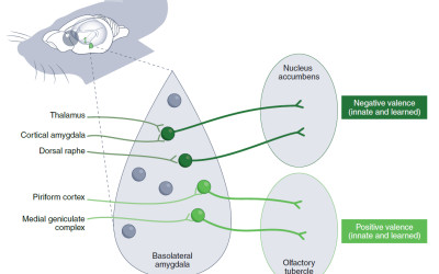
The basolateral amygdala (BLA) mediates associative learning for both fear and reward. Accumulating evidence supports the notion that different BLA projections distinctly alter motivated behavior, including projections to the nucleus accumbens (NAc), medial aspect of the central amygdala (CeM), and ventral hippocampus (vHPC). Although there is consensus regarding the existence of distinct subsets of BLA neurons encoding positive or negative valence, controversy remains regarding the anatomical arrangement of these populations. First, we map the location of more than 1,000 neurons distributed across the BLA and recorded during a Pavlovian discrimination task. Next, we determine the location of projection-defined neurons labeled with retrograde tracers and use CLARITY to reveal the axonal path in 3-dimensional space. Finally, we examine the local influence of each projection-defined populations within the BLA. Understanding the functional and topographical organization of circuits underlying valence assignment could reveal fundamental principles about emotional processing.


06/2017 | Nat Neurosci
Amygdala inputs to prefrontal cortex guide behavior amid conflicting cues of reward and punishment.
Burgos-Robles A, Kimchi EY, Izadmehr EM, Porzenheim MJ, Ramos-Guasp WA, Nieh EH, Felix-Ortiz AC, Namburi P, Leppla CA, Presbrey KN, Anandalingam KK, Pagan-Rivera PA, Anahtar M, Beyeler A, Tye KM
doi: 10.1038/nn.4553

Abstract:
Orchestrating appropriate behavioral responses in the face of competing signals that predict either rewards or threats in the environment is crucial for survival. The basolateral nucleus of the amygdala (BLA) and prelimbic (PL) medial prefrontal cortex have been implicated in reward-seeking and fear-related responses, but how information flows between these reciprocally connected structures to coordinate behavior is unknown. We recorded neuronal activity from the BLA and PL while rats performed a task wherein competing shock- and sucrose-predictive cues were simultaneously presented. The correlated firing primarily displayed a BLA-->PL directionality during the shock-associated cue. Furthermore, BLA neurons optogenetically identified as projecting to PL more accurately predicted behavioral responses during competition than unidentified BLA neurons. Finally photostimulation of the BLA-->PL projection increased freezing, whereas both chemogenetic and optogenetic inhibition reduced freezing. Therefore, the BLA-->PL circuit is critical in governing the selection of behavioral responses in the face of competing signals.


04/11/2016 | Science
Parsing reward from aversion.
Beyeler A


20/04/2016 | Neuron
Divergent Routing of Positive and Negative Information from the Amygdala during Memory Retrieval.
Beyeler A, Namburi P, Glober GF, Simonnet C, Calhoon GG, Conyers GF, Luck R, Wildes CP, Tye KM
doi: 10.1016/j.neuron.2016.03.004

Abstract:
Although the basolateral amygdala (BLA) is known to play a critical role in the formation of memories of both positive and negative valence, the coding and routing of valence-related information is poorly understood. Here, we recorded BLA neurons during the retrieval of associative memories and used optogenetic-mediated phototagging to identify populations of neurons that synapse in the nucleus accumbens (NAc), the central amygdala (CeA), or ventral hippocampus (vHPC). We found that despite heterogeneous neural responses within each population, the proportions of BLA-NAc neurons excited by reward predictive cues and of BLA-CeA neurons excited by aversion predictive cues were higher than within the entire BLA. Although the BLA-vHPC projection is known to drive behaviors of innate negative valence, these neurons did not preferentially code for learned negative valence. Together, these findings suggest that valence encoding in the BLA is at least partially mediated via divergent activity of anatomically defined neural populations.


30/04/2015 | Nature
A circuit mechanism for differentiating positive and negative associations.
Namburi P*, Beyeler A*, Yorozu S, Calhoon GG, Halbert SA, Wichmann R, Holden SS, Mertens KL, Anahtar M, Felix-Ortiz AC, Wickersham IR, Gray JM, Tye KM
doi: 10.1038/nature14366

Abstract:
The ability to differentiate stimuli predicting positive or negative outcomes is critical for survival, and perturbations of emotional processing underlie many psychiatric disease states. Synaptic plasticity in the basolateral amygdala complex (BLA) mediates the acquisition of associative memories, both positive and negative. Different populations of BLA neurons may encode fearful or rewarding associations, but the identifying features of these populations and the synaptic mechanisms of differentiating positive and negative emotional valence have remained unknown. Here we show that BLA neurons projecting to the nucleus accumbens (NAc projectors) or the centromedial amygdala (CeM projectors) undergo opposing synaptic changes following fear or reward conditioning. We find that photostimulation of NAc projectors supports positive reinforcement while photostimulation of CeM projectors mediates negative reinforcement. Photoinhibition of CeM projectors impairs fear conditioning and enhances reward conditioning. We characterize these functionally distinct neuronal populations by comparing their electrophysiological, morphological and genetic features. Overall, we provide a mechanistic explanation for the representation of positive and negative associations within the amygdala.


2014 | prog mol biol transl sci
Deciphering memory function with optogenetics.
Beyeler A, Eckhardt CA, Tye KM
doi: 10.1016/B978-0-12-420170-5.00012-X

Abstract:
Optogenetics has accelerated the field of neuroscience by overcoming many of the spatial, genetic, and temporal limitations of previous techniques to control neural activity. The study of learning and memory has profoundly benefited from these tools mainly from their use in rodents. New insights have been made regarding the involvement of specific cell types or populations of synapses in the acquisition, consolidation, and retrieval of memories. The cellular specificity and temporal precision of optogenetic manipulations have also shown to be useful to study synaptic mechanisms supporting learning and memory including long-term synaptic plasticity. Recently, new light-sensitive molecules have been developed to control intracellular pathways or gene expression, which promise to enhance our understanding of the molecular mechanism of memory function.


21/08/2013 | Neuron
BLA to vHPC inputs modulate anxiety-related behaviors.
Felix-Ortiz AC*, Beyeler A*, Seo C, Leppla CA, Wildes CP, Tye KM
doi: 10.1016/j.neuron.2013.06.016

Abstract:
The basolateral amygdala (BLA) and ventral hippocampus (vHPC) have both been implicated in mediating anxiety-related behaviors, but the functional contribution of BLA inputs to the vHPC has never been directly investigated. Here we show that activation of BLA-vHPC synapses acutely and robustly increased anxiety-related behaviors, while inhibition of BLA-vHPC synapses decreased anxiety-related behaviors. We combined optogenetic approaches with in vivo pharmacological manipulations and ex vivo whole-cell patch-clamp recordings to dissect the local circuit mechanisms, demonstrating that activation of BLA terminals in the vHPC provided monosynaptic, glutamatergic inputs to vHPC pyramidal neurons. Furthermore, BLA inputs exerted polysynaptic, inhibitory effects mediated by local interneurons in the vHPC that may serve to balance the circuit locally. These data establish a role for BLA-vHPC synapses in bidirectionally controlling anxiety-related behaviors in an immediate, yet reversible, manner and a model for the local circuit mechanism of BLA inputs in the vHPC.


2013 | PLoS ONE
Recruitment of Perisomatic Inhibition during Spontaneous Hippocampal Activity
Beyeler A, Retailleau A, Molter C, Mehidi A, Szabadics J, Leinekugel X
doi: 10.1371/journal.pone.0066509

Abstract:
It was recently shown that perisomatic GABAergic inhibitory postsynaptic potentials (IPSPs) originating from basket and chandelier cells can be recorded as population IPSPs from the hippocampal pyramidal layer using extracellular electrodes (eIPSPs). Taking advantage of this approach, we have investigated the recruitment of perisomatic inhibition during spontaneous hippocampal activity in vitro. Combining intracellular and extracellular recordings from pyramidal cells and interneurons, we confirm that inhibitory signals generated by basket cells can be recorded extracellularly, but our results suggest that, during spontaneous activity, eIPSPs are mostly confined to the CA3 rather than CA1 region. CA3 eIPSPs produced the powerful time-locked inhibition of multi-unit activity expected from perisomatic inhibition. Analysis of the temporal dynamics of spike discharges relative to eIPSPs suggests significant but moderate recruitment of excitatory and inhibitory neurons within the CA3 network on a 10 ms time scale, within which neurons recruit each other through recurrent collaterals and trigger powerful feedback inhibition. Such quantified parameters of neuronal interactions in the hippocampal network may serve as a basis for future characterisation of pathological conditions potentially affecting the interactions between excitation and inhibition in this circuit.


Abstract:
During frog metamorphosis, the vestibular sensory system remains unchanged, while spinal motor networks undergo a massive restructuring associated with the transition from the larval to adult biomechanical system. We investigated in Xenopus laevis the impact of a pre- (tadpole stage) or post-metamorphosis (juvenile stage) unilateral labyrinthectomy (UL) on young adult swimming performance and underlying spinal locomotor circuitry. The acute disruptive effects on locomotion were similar in both tadpoles and juvenile frogs. However, animals that had metamorphosed with a preceding UL expressed restored swimming behavior at the juvenile stage, whereas animals lesioned after metamorphosis never recovered. Whilst kinematic and electrophysiological analyses of the propulsive system showed no significant differences in either juvenile group, a 3D biomechanical simulation suggested that an asymmetry in the dynamic control of posture during swimming could account for the behavioral restoration observed in animals that had been labyrinthectomized before metamorphosis. This hypothesis was subsequently supported by in vivo electromyography during free swimming and in vitro recordings from isolated brainstem/spinal cord preparations. Specifically, animals lesioned prior to metamorphosis at the larval stage exhibited an asymmetrical propulsion/posture coupling as a post-metamorphic young adult. This developmental alteration was accompanied by an ipsilesional decrease in propriospinal coordination that is normally established in strict left-right symmetry during metamorphosis in order to synchronize dorsal trunk muscle contractions with bilateral hindlimb extensions in the swimming adult. Our data thus suggest that a disequilibrium in descending vestibulospinal information during Xenopus metamorphosis leads to an altered assembly of adult spinal locomotor circuitry. This in turn enables an adaptive compensation for the dynamic postural asymmetry induced by the vestibular imbalance and the restoration of functionally-effective behavior.


05/2012 | J Physiol Paris
Neural circuits underlying the generation of theta oscillations.
Pignatelli M, Beyeler A, Leinekugel X
doi: 10.1016/j.jphysparis.2011.09.007

Abstract:
Theta oscillations represent the neural network configuration underlying active awake behavior and paradoxical sleep. This major EEG pattern has been extensively studied, from physiological to anatomical levels, for more than half a century. Nevertheless the cellular and network mechanisms accountable for the theta generation are still not fully understood. This review synthesizes the current knowledge on the circuitry involved in the generation of theta oscillations, from the hippocampus to extra hippocampal structures such as septal complex, entorhinal cortex and pedunculopontine tegmentum, a main trigger of theta state through direct and indirect projections to the septal complex. We conclude with a short overview of the perspectives offered by technical advances for deciphering more precisely the different neural components underlying the emergence of theta oscillations.


Abstract:
Serotonin2C (5-HT(2C)) receptors act in the basal ganglia, a group of sub-cortical structures involved in motor behavior, where they are thought to modulate oral activity and participate in iatrogenic motor side-effects in Parkinson's disease and Schizophrenia. Whether abnormal movements initiated by 5-HT(2C) receptors are directly consequent to dysfunctions of the motor circuit is uncertain. In the present study, we combined behavioral, immunohistochemical and extracellular single-cell recordings approaches in rats to investigate the effect of the 5-HT(2C) agonist Ro-60-0175 respectively on orofacial dyskinesia, the expression of the marker of neuronal activity c-Fos in basal ganglia and the electrophysiological activity of substantia nigra pars reticulata (SNr) neuron connected to the orofacial motor cortex (OfMC) or the medial prefrontal cortex (mPFC). The results show that Ro-60-0175 (1 mg/kg) caused bouts of orofacial movements that were suppressed by the 5-HT(2C) antagonist SB-243213 (1 mg/kg). Ro-60-0175 (0.3, 1, 3 mg/kg) dose-dependently enhanced Fos expression in the striatum and the nucleus accumbens. At the highest dose, it enhanced Fos expression in the subthalamic nucleus, the SNr and the entopeduncular nucleus but not in the external globus pallidus. However, the effect of Ro-60-0175 was mainly associated with associative/limbic regions of basal ganglia whereas subregions of basal ganglia corresponding to sensorimotor territories were devoid of Fos labeling. Ro-60-0175 (1-3 mg/kg) did not affect the electrophysiological activity of SNr neurons connected to the OfMC nor their excitatory-inhibitory-excitatory responses to the OfMC electrical stimulation. Conversely, Ro-60-0175 (1 mg/kg) enhanced the late excitatory response of SNr neurons evoked by the mPFC electrical stimulation. These results suggest that oral dyskinesia induced by 5-HT(2C) agonists are not restricted to aberrant signalling in the orofacial motor circuit and demonstrate discrete modifications in associative territories.


Abstract:
Anuran metamorphosis includes a complete remodeling of the animal's biomechanical apparatus, requiring a corresponding functional reorganization of underlying central neural circuitry. This involves changes that must occur in the coordination between the motor outputs of different spinal segments to harmonize locomotor and postural functions as the limbs grow and the tail regresses. In premetamorphic Xenopus laevis tadpoles, axial motor output drives rostrocaudally propagating segmental myotomal contractions that generate propulsive body undulations. During metamorphosis, the anterior axial musculature of the tadpole progressively evolves into dorsal muscles in the postmetamorphic froglet in which some of these back muscles lose their implicit locomotor function to serve exclusively in postural control in the adult. To understand how locomotor and postural systems interact during locomotion in juvenile Xenopus, we have investigated the coordination between postural back and hindlimb muscle activity during free forward swimming. Axial/dorsal muscles, which contract in bilateral alternation during undulatory swimming in premetamorphic tadpoles, change their left-right coordination to become activated in phase with bilaterally synchronous hindlimb extensions in locomoting juveniles. Based on in vitro electrophysiological experiments as well as specific spinal lesions in vivo, a spinal cord region was delimited in which propriospinal interactions are directly responsible for the coordination between leg and back muscle contractions. Our findings therefore indicate that dynamic postural adjustments during adult Xenopus locomotion are mediated by local intraspinal pathways through which the lumbar generator for hindlimb propulsive kicking provides caudorostral commands to thoracic spinal circuitry controlling the dorsal trunk musculature.