IF du Neurocentre
IF1234567891011121314151617181920253035404550
Nombre00062656011001632201213311
%00083878022002843302324422

83 publications


12/09/2024 | adv healthc mater
Dye-Based Fluorescent Organic Nanoparticles, New Promising Tools for Optogenetics.
Lesas J, Bienvenu TCM, Kurek E, Verlhac JB, Grivet Z, Têtu M, Girard D, Lanore F, Blanchard-Desce M, Herry C, Daniel J, Dejean C
doi: 10.1002/adhm.202402132

Abstract:
Dye-based fluorescent organic nanoparticles are a specific class of nanoparticles obtained by nanoprecipitation in water of pure dyes only. While the photophysical and colloidal properties of the nanoparticles strongly depend on the nature of the aggregated dyes, their excellent brightness in the visible and in the near infrared make these nanoparticles a unique and versatile platform for in vivo application. This article examines the promising utilization of these nanoparticles for in vivo optogenetics applications. Their photophysical properties as well as their biocompatibility and their capacity to activate Chrimson opsin in vivo through the fluorescence reabsorption process are demonstrated. Additionally, an illustrative example of employing these nanoparticles in fear reduction in mice through closed-loop stimulation is presented. Through an optogenetic methodology, the nanoparticles demonstrate an ability to selectively manipulate neurons implicated in the fear response and diminish the latter. Dye-based fluorescent organic nanoparticles represent a promising and innovative strategy for optogenetic applications, holding substantial potential in the domain of translational neuroscience. This work paves the way for novel therapeutic modalities for neurological and neuropsychiatric disorders.


Abstract:
The dynamic suppression of threat-related behavior as a function of environmental constraint is critical for survival in mammals, yet the neurobiological underpinnings remain largely unknown. In this issue of Neuron, Wang et al.(1) identified prefrontal dynorphin-expressing neurons as key elements for tracking threat-related behavioral states and regulating fear suppression.


05/03/2024 | J Neurol Neurosurg Psychiatry
Resective epilepsy surgery and its impact on depression in adults: a systematic review, meta-analysis, and implications for future research.
Hernandez Poblete N, Gay F, Salvo F, Micoulaud-Franchi JA, Bienvenu T, Coelho J, Aupy J
doi: 10.1136/jnnp-2023-333073

Abstract:
BACKGROUND: How epilepsy surgery influences the bidirectional relationship of epilepsy and depression remains poorly defined. METHOD: For a better understanding of this question, we conducted a systematic review and meta-analysis of risk ratio on depression prevalence before and after epilepsy surgery, using Preferred Reporting Items for Systematic reviews and Meta-Analyses guidelines. Three databases were comprehensively screened for all studies assessing depression before and after resective surgery in adult epileptic patients until 8 October 2022. Studies were included if depression was assessed before and after epilepsy surgery regardless of the time of follow-up. A total of 1917 studies were screened for eligibility and 91 full-texts up for inclusion; 35 studies were finally included, 25 studies and 2563 patients were included in main meta-analysis and 10 for exploratory analysis. Risk of bias was assessed using Risk Of Bias In Non-randomised Studies - of Interventions (ROBINS-I) from Cochrane. To derive the pooled depression rates before and after surgery, a meta-analysis with inversed-variance was performed using random-effects logistic models with Peto's correction and a 95% CI. Heterogeneity was assessed with Cochran's Q-test along with its derived measure of inconsistency I(2). RESULTS: Overall, the depression rates before and after resective epilepsy surgery were 0.70 (0.53 to 0.91) 95% CI, suggesting that the rate of depression at last follow-up evaluation tends to decrease after Resective Epilepsy Surgery (RES). Subgroup analysis suggest a positive long-term effect appears with a significant lower rates of depression already 6 months (0.61 (0.38 to 0.98)), after surgery which is maintained over time after 1 year (0.53 (0.31 to 0.90)), and after 2 years (0.62 (0.42 to 0.92)). CONCLUSION: This important finding should be taken in consideration before resective surgery for drug-resistant epilepsies. However, prospective studies should be conducted to characterise which patient, at the individual level, might be at risk of de novo or worsening of depression. PROSPERO REGISTRATION NUMBER: CRD42022355386.


29/02/2024 | Sci Rep
Genetic labeling of embryonically-born dentate granule neurons in young mice using the Penk(Cre) mouse line.
Mortessagne P, Cartier E, Balia M, Fevre M, Corailler F, Herry C, Abrous DN, Battefeld A, Pacary E
doi: 10.1038/s41598-024-55299-9

Abstract:
The dentate gyrus (DG) of the hippocampus is a mosaic of dentate granule neurons (DGNs) accumulated throughout life. While many studies focused on the morpho-functional properties of adult-born DGNs, much less is known about DGNs generated during development, and in particular those born during embryogenesis. One of the main reasons for this gap is the lack of methods available to specifically label and manipulate embryonically-born DGNs. Here, we have assessed the relevance of the Penk(Cre) mouse line as a genetic model to target this embryonically-born population. In young animals, Penk(Cre) expression allows to tag neurons in the DG with positional, morphological and electrophysiological properties characteristic of DGNs born during the embryonic period. In addition, Penk(Cre)+ cells in the DG are distributed in both blades along the entire septo-temporal axis. This model thus offers new possibilities to explore the functions of this underexplored population of embryonically-born DGNs.


14/12/2023 | Nat Commun
CA3 hippocampal synaptic plasticity supports ripple physiology during memory consolidation.
El Oussini H, Zhang CL, Francois U, Castelli C, Lampin-Saint-Amaux A, Lepleux M, Molle P, Velez L, Dejean C, Lanore F, Herry C, Choquet D, Humeau Y
doi: 10.1038/s41467-023-42969-x

Abstract:
The consolidation of recent memories depends on memory replays, also called ripples, generated within the hippocampus during slow-wave sleep, and whose inactivation leads to memory impairment. For now, the mobilisation, localisation and importance of synaptic plasticity events associated to ripples are largely unknown. To tackle this question, we used cell surface AMPAR immobilisation to block post-synaptic LTP within the hippocampal region of male mice during a spatial memory task, and show that: 1- hippocampal synaptic plasticity is engaged during consolidation, but is dispensable during encoding or retrieval. 2- Plasticity blockade during sleep results in apparent forgetting of the encoded rule. 3- In vivo ripple recordings show a strong effect of AMPAR immobilisation when a rule has been recently encoded. 4- In situ investigation suggests that plasticity at CA3-CA3 recurrent synapses supports ripple generation. We thus propose that post-synaptic AMPAR mobility at CA3 recurrent synapses is necessary for ripple-dependent rule consolidation.


30/10/2023 | Nat Neurosci
Prefrontal circuits encode both general danger and specific threat representations.
Martin-Fernandez M, Menegolla AP, Lopez-Fernandez G, Winke N, Jercog D, Kim HR, Girard D, Dejean C, Herry C
doi: 10.1038/s41593-023-01472-8

Abstract:
Behavioral adaptation to potential threats requires both a global representation of danger to prepare the organism to react in a timely manner but also the identification of specific threatening situations to select the appropriate behavioral responses. The prefrontal cortex is known to control threat-related behaviors, yet it is unknown whether it encodes global defensive states and/or the identity of specific threatening encounters. Using a new behavioral paradigm that exposes mice to different threatening situations, we show that the dorsomedial prefrontal cortex (dmPFC) encodes a general representation of danger while simultaneously encoding a specific neuronal representation of each threat. Importantly, the global representation of danger persisted in error trials that instead lacked specific threat identity representations. Consistently, optogenetic prefrontal inhibition impaired overall behavioral performance and discrimination of different threatening situations without any bias toward active or passive behaviors. Together, these data indicate that the prefrontal cortex encodes both a global representation of danger and specific representations of threat identity to control the selection of defensive behaviors.


02/10/2023 | Sci Rep
Integrating operant behavior and fiber photometry with the open-source python library Pyfiber.
Conlisk D, Ceau M, Fiancette JF, Winke N, Darmagnac E, Herry C, Deroche-Gamonet V
doi: 10.1038/s41598-023-43565-1

Abstract:
Despite the popularity of fiber photometry (FP), its integration with operant behavior paradigms is progressing slowly. This can be attributed to the complex protocols in operant behavior - resulting in a combination of diverse non-predictable behavioral responses and scheduled events, thereby complicating data analysis. To overcome this, we developed Pyfiber, an open-source python library which facilitates the merge of FP with operant behavior by relating changes in fluorescent signals within a neuronal population to behavioral responses and events. Pyfiber helps to 1. Extract events and responses that occur in operant behavior, 2. Extract and process the FP signals, 3. Select events of interest and align them to the corresponding FP signals, 4. Apply appropriate signal normalization and analysis according to the type of events, 5. Run analysis on multiple individuals and sessions, 6. Collect results in an easily readable format. Pyfiber is suitable for use with many different fluorescent sensors and operant behavior protocols. It was developed using Doric lenses FP systems and Imetronic behavioral systems, but it possesses the capability to process data from alternative systems. This work sets a solid foundation for analyzing the relationship between different dimensions of complex behavioral paradigms with fluorescent signals from brain regions of interest.


18/01/2023 | sci adv
Disynaptic specificity of serial information flow for conditioned fear.
Massi L, Hagihara KM, Courtin J, Hinz J, Muller C, Fustinana MS, Xu C, Karalis N, Luthi A
doi: 10.1126/sciadv.abq1637

Abstract:
Memory encoding and retrieval rely on specific interactions across multiple brain areas. Although connections between individual brain areas have been extensively studied, the anatomical and functional specificity of neuronal circuit organization underlying information transfer across multiple brain areas remains unclear. Here, we combine transsynaptic viral tracing, optogenetic manipulations, and calcium dynamics recordings to dissect the multisynaptic functional connectivity of the amygdala. We identify a distinct basolateral amygdala (BLA) subpopulation that connects disynaptically to the periaqueductal gray (PAG) via the central amygdala (CeA). This disynaptic pathway serves as a core circuit element necessary for the learning and expression of conditioned fear and exhibits learning-related plasticity. Together, our findings demonstrate the utility of multisynaptic approaches for functional circuit analysis and indicate that disynaptic specificity may be a general feature of neuronal circuit organization.


2023 | Front Cell Neurosci
Axo-axonic cells in neuropsychiatric disorders: a systematic review.
Vivien J, El Azraoui A, Lheraux C, Lanore F, Aouizerate B, Herry C, Humeau Y, Bienvenu TCM

Abstract:
Imbalance between excitation and inhibition in the cerebral cortex is one of the main theories in neuropsychiatric disorder pathophysiology. Cortical inhibition is finely regulated by a variety of highly specialized GABAergic interneuron types, which are thought to organize neural network activities. Among interneurons, axo-axonic cells are unique in making synapses with the axon initial segment of pyramidal neurons. Alterations of axo-axonic cells have been proposed to be implicated in disorders including epilepsy, schizophrenia and autism spectrum disorder. However, evidence for the alteration of axo-axonic cells in disease has only been examined in narrative reviews. By performing a systematic review of studies investigating axo-axonic cells and axo-axonic communication in epilepsy, schizophrenia and autism spectrum disorder, we outline convergent findings and discrepancies in the literature. Overall, the implication of axo-axonic cells in neuropsychiatric disorders might have been overstated. Additional work is needed to assess initial, mostly indirect findings, and to unravel how defects in axo-axonic cells translates to cortical dysregulation and, in turn, to pathological states.


29/07/2022 | sci adv
Switch of serotonergic descending inhibition into facilitation by a spinal chloride imbalance in neuropathic pain.
Aby F, Lorenzo LE, Grivet Z, Bouali-Benazzouz R, Martin H, Valerio S, Whitestone S, Isabel D, Idi W, Bouchatta O, De Deurwaerdere P, Godin AG, Herry C, Fioramonti X, Landry M, De Koninck Y, Fossat P
doi: 10.1126/sciadv.abo0689

Abstract:
Descending control from the brain to the spinal cord shapes our pain experience, ranging from powerful analgesia to extreme sensitivity. Increasing evidence from both preclinical and clinical studies points to an imbalance toward descending facilitation as a substrate of pathological pain, but the underlying mechanisms remain unknown. We used an optogenetic approach to manipulate serotonin (5-HT) neurons of the nucleus raphe magnus that project to the dorsal horn of the spinal cord. We found that 5-HT neurons exert an analgesic action in naïve mice that becomes proalgesic in an experimental model of neuropathic pain. We show that spinal KCC2 hypofunction turns this descending inhibitory control into paradoxical facilitation; KCC2 enhancers restored 5-HT-mediated descending inhibition and analgesia. Last, combining selective serotonin reuptake inhibitors (SSRIs) with a KCC2 enhancer yields effective analgesia against nerve injury-induced pain hypersensitivity. This uncovers a previously unidentified therapeutic path for SSRIs against neuropathic pain.


06/07/2022 | Curr Opin Neurobiol
Decoding defensive systems.
Herry C, Jercog D
doi: 10.1016/j.conb.2022.102600

Abstract:
Our understanding of the neuronal circuits and mechanisms of defensive systems has been primarily dominated by studies focusing on the contribution of individual cells in the processing of threat-predictive cues, defensive responses, the extinction of such responses and the contextual modulation of threat-related behavior. These studies have been key in establishing threat-related circuits and mechanisms. Yet, they fall short in answering long-standing questions related to the integrative processing of distinct threatening cues, behavioral states induced by threat-related events, or the bridging from sensory processing of threat-related cues to specific defensive responses. Recent conceptual and technical developments has allowed the monitoring of large populations of neurons, which in addition to advanced analytic tools, have improved our understanding of how collective neuronal activity supports threat-related behaviors. In this review, we discuss the current knowledge of neuronal population codes within threat-related networks, in the context of aversive motivated behavior and the study of defensive systems.


10/01/2022 | Nat Commun
Vascular and blood-brain barrier-related changes underlie stress responses and resilience in female mice and depression in human tissue.
Dion-Albert L, Cadoret A, Doney E, Kaufmann FN, Dudek KA, Daigle B, Parise LF, Cathomas F, Samba N, Hudson N, Lebel M, Campbell M, Turecki G, Mechawar N, Menard C
doi: 10.1038/s41467-021-27604-x

Abstract:
Prevalence, symptoms, and treatment of depression suggest that major depressive disorders (MDD) present sex differences. Social stress-induced neurovascular pathology is associated with depressive symptoms in male mice; however, this association is unclear in females. Here, we report that chronic social and subchronic variable stress promotes blood-brain barrier (BBB) alterations in mood-related brain regions of female mice. Targeted disruption of the BBB in the female prefrontal cortex (PFC) induces anxiety- and depression-like behaviours. By comparing the endothelium cell-specific transcriptomic profiling of the mouse male and female PFC, we identify several pathways and genes involved in maladaptive stress responses and resilience to stress. Furthermore, we confirm that the BBB in the PFC of stressed female mice is leaky. Then, we identify circulating vascular biomarkers of chronic stress, such as soluble E-selectin. Similar changes in circulating soluble E-selectin, BBB gene expression and morphology can be found in blood serum and postmortem brain samples from women diagnosed with MDD. Altogether, we propose that BBB dysfunction plays an important role in modulating stress responses in female mice and possibly MDD.


07/01/2022 | Science
A neuronal mechanism for motivational control of behavior.
Courtin J, Bitterman Y, Muller S, Hinz J, Hagihara KM, Muller C, Luthi A
doi: 10.1126/science.abg7277

Abstract:
Acting to achieve goals depends on the ability to motivate specific behaviors based on their predicted consequences given an individual's internal state. However, the underlying neuronal mechanisms that encode and maintain such specific motivational control of behavior are poorly understood. Here, we used Ca(2+) imaging and optogenetic manipulations in the basolateral amygdala of freely moving mice performing noncued, self-paced instrumental goal-directed actions to receive and consume rewards. We found that distinct neuronal activity patterns sequentially represent the entire action-consumption behavioral sequence. Whereas action-associated patterns integrated the identity, value, and expectancy of pursued goals, consumption-associated patterns reflected the identity and value of experienced outcomes. Thus, the interplay between these patterns allows the maintenance of specific motivational states necessary to adaptively direct behavior toward prospective rewards.


Abstract:
Background: Pathological anxiety is responsible for major functional impairments and resistance to conventional treatments in anxiety disorders (ADs), posttraumatic stress disorder (PTSD) and major depressive disorder (MDD). Focal neuromodulation therapies such as transcranial magnetic stimulation (TMS), transcranial direct current stimulation (tDCS) and deep brain stimulation (DBS) are being developed to treat those disorders. Methods: We performed a dimensional systematic review and meta-analysis to assess the evidence of the efficacy of TMS, tDCS and DBS in reducing anxiety symptoms across ADs, PTSD and MDD. Reports were identified through systematic searches in PubMed/Medline, Scopus and Cochrane library (inception to November 2020), followed by review according to the PRISMA guidelines. Controlled clinical trials examining the effectiveness of brain stimulation techniques on generic anxiety symptoms in patients with ADs, PTSD or MDD were selected. Results: Nineteen studies (RCTs) met inclusion criteria, which included 589 participants. Overall, focal brain activity modulation interventions were associated with greater reduction of anxiety levels than controls [SMD: -0.56 (95% CI, -0.93 to-0.20, I (2) = 77%]. Subgroup analyses revealed positive effects for TMS across disorders, and of focal neuromodulation in generalized anxiety disorder and PTSD. Rates of clinical responses and remission were higher in the active conditions. However, the risk of bias was high in most studies. Conclusions: There is moderate quality evidence for the efficacy of neuromodulation in treating pathological anxiety. Systematic Review Registration: https://www.crd.york.ac.uk/prospero/display_record.php?RecordID=233084, identifier: PROSPERO CRD42021233084. It was submitted on January 29th, 2021, and registered on March 1st, 2021. No amendment was made to the recorded protocol. A change was applied for the subgroup analyses based on target brain regions, we added the putative nature (excitatory/inhibitory) of brain activity modulation.


15/09/2021 | Mol Psychiatry
The temporal origin of dentate granule neurons dictates their role in spatial memory.
Masachs N, Charrier V, Farrugia F, Lemaire V, Blin N, Mazier W, Tronel S, Montaron MF, Ge S, Marsicano G, Cota D, Deroche-Gamonet V, Herry C, Abrous DN
doi: 10.1038/s41380-021-01276-x

Abstract:
The dentate gyrus is one of the only brain regions that continues its development after birth in rodents. Adolescence is a very sensitive period during which cognitive competences are programmed. We investigated the role of dentate granule neurons (DGNs) born during adolescence in spatial memory and compared them with those generated earlier in life (in embryos or neonates) or during adulthood by combining functional imaging, retroviral and optogenetic tools to tag and silence DGNs. By imaging DGNs expressing Zif268, a proxy for neuronal activity, we found that neurons generated in adolescent rats (and not embryos or neonates) are transiently involved in spatial memory processing. In contrast, adult-generated DGNs are recruited at a later time point when animals are older. A causal relationship between the temporal origin of DGNs and spatial memory was confirmed by silencing DGNs in behaving animals. Our results demonstrate that the emergence of spatial memory depends on neurons born during adolescence, a function later assumed by neurons generated during adulthood.


14/09/2021 | Glia
N-3 PUFA deficiency disrupts oligodendrocyte maturation and myelin integrity during brain development.
Leyrolle Q, Decoeur F, Dejean C, Briere G, Leon S, Bakoyiannis I, Baroux E, Sterley TL, Bosch-Bouju C, Morel L, Amadieu C, Lecours C, St-Pierre MK, Bordeleau M, De Smedt-Peyrusse V, Sere A, Schwendimann L, Gregoire S, Bretillon L, Acar N, Joffre C, Ferreira G, Uricaru R, Thebault P, Gressens P, Tremblay ME, Laye S, Nadjar A
doi: 10.1002/glia.24088

Abstract:
Westernization of dietary habits has led to a progressive reduction in dietary intake of n-3 polyunsaturated fatty acids (n-3 PUFAs). Low maternal intake of n-3 PUFAs has been linked to neurodevelopmental disorders, conditions in which myelination processes are abnormal, leading to defects in brain functional connectivity. Only little is known about the role of n-3 PUFAs in oligodendrocyte physiology and white matter development. Here, we show that lifelong n-3 PUFA deficiency disrupts oligodendrocytes maturation and myelination processes during the postnatal period in mice. This has long-term deleterious consequences on white matter organization and hippocampus-prefrontal functional connectivity in adults, associated with cognitive and emotional disorders. Promoting developmental myelination with clemastine, a first-generation histamine antagonist and enhancer of oligodendrocyte precursor cell differentiation, rescues memory deficits in n-3 PUFA deficient animals. Our findings identify a novel mechanism through which n-3 PUFA deficiency alters brain functions by disrupting oligodendrocyte maturation and brain myelination during the neurodevelopmental period.


14/07/2021 | Nature
Dynamical prefrontal population coding during defensive behaviours.
Jercog D, Winke N, Sung K, Fernandez MM, Francioni C, Rajot D, Courtin J, Chaudun F, Jercog PE, Valerio S, Herry C
doi: 10.1038/s41586-021-03726-6

Abstract:
Coping with threatening situations requires both identifying stimuli that predict danger and selecting adaptive behavioural responses to survive(1). The dorsomedial prefrontal cortex (dmPFC) is a critical structure that is involved in the regulation of threat-related behaviour(2-4). However, it is unclear how threat-predicting stimuli and defensive behaviours are associated within prefrontal networks to successfully drive adaptive responses. Here we used a combination of extracellular recordings, neuronal decoding approaches, pharmacological and optogenetic manipulations to show that, in mice, threat representations and the initiation of avoidance behaviour are dynamically encoded in the overall population activity of dmPFC neurons. Our data indicate that although dmPFC population activity at stimulus onset encodes sustained threat representations driven by the amygdala, it does not predict action outcome. By contrast, transient dmPFC population activity before the initiation of action reliably predicts avoided from non-avoided trials. Accordingly, optogenetic inhibition of prefrontal activity constrained the selection of adaptive defensive responses in a time-dependent manner. These results reveal that the adaptive selection of defensive responses relies on a dynamic process of information linking threats with defensive actions, unfolding within prefrontal networks.


06/07/2021 | Nat Commun
Central amygdala micro-circuits mediate fear extinction.
Whittle N, Fadok J, MacPherson KP, Nguyen R, Botta P, Wolff SBE, Müller C, Herry C, Tovote P, Holmes A, Singewald N, Lüthi A, Ciocchi S
doi: 10.1038/s41467-021-24068-x

Abstract:
Fear extinction is an adaptive process whereby defensive responses are attenuated following repeated experience of prior fear-related stimuli without harm. The formation of extinction memories involves interactions between various corticolimbic structures, resulting in reduced central amygdala (CEA) output. Recent studies show, however, the CEA is not merely an output relay of fear responses but contains multiple neuronal subpopulations that interact to calibrate levels of fear responding. Here, by integrating behavioural, in vivo electrophysiological, anatomical and optogenetic approaches in mice we demonstrate that fear extinction produces reversible, stimulus- and context-specific changes in neuronal responses to conditioned stimuli in functionally and genetically defined cell types in the lateral (CEl) and medial (CEm) CEA. Moreover, we show these alterations are absent when extinction is deficient and that selective silencing of protein kinase C delta-expressing (PKCδ) CEl neurons impairs fear extinction. Our findings identify CEA inhibitory microcircuits that act as critical elements within the brain networks mediating fear extinction.


Abstract:
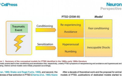
Translational research on post-traumatic stress disorder (PTSD) has produced limited improvements in clinical practice. Fear conditioning (FC) is one of the dominant animal models of PTSD. In fact, FC is used in many different ways to model PTSD. The variety of FC-based models is ill defined, creating confusion and conceptual vagueness, which in turn impedes translation into the clinic. This article takes a historical and conceptual approach to provide a comprehensive picture of current research and help reorient the research focus. This work historically reviews the variety of models that have emerged from the initial association of PTSD with FC, highlighting conceptual pitfalls that have limited the translation of animal research into clinical advances. We then provide some guidance on how future translational research could benefit from conceptual and technological improvements to translate basic findings in patients. This objective will require transdisciplinary approaches and should involve physicians, engineers, philosophers, and neuroscientists.


10/05/2021 | Nat Commun
Breathing-driven prefrontal oscillations regulate maintenance of conditioned-fear evoked freezing independently of initiation.
Bagur S, Lefort JM, Lacroix MM, de Lavilléon G, Herry C, Chouvaeff M, Billand C, Geoffroy H, Benchenane K
doi: 10.1038/s41467-021-22798-6

Abstract:
Brain-body interactions are thought to be essential in emotions but their physiological basis remains poorly understood. In mice, regular 4 Hz breathing appears during freezing after cue-fear conditioning. Here we show that the olfactory bulb (OB) transmits this rhythm to the dorsomedial prefrontal cortex (dmPFC) where it organizes neural activity. Reduction of the respiratory-related 4 Hz oscillation, via bulbectomy or optogenetic perturbation of the OB, reduces freezing. Behavioural modelling shows that this is due to a specific reduction in freezing maintenance without impacting its initiation, thus dissociating these two phenomena. dmPFC LFP and firing patterns support the region's specific function in freezing maintenance. In particular, population analysis reveals that network activity tracks 4 Hz power dynamics during freezing and reaches a stable state at 4 Hz peak that lasts until freezing termination. These results provide a potential mechanism and a functional role for bodily feedback in emotions and therefore shed light on the historical James-Cannon debate.


11/2019 | Nat Neurosci
Adaptive disinhibitory gating by VIP interneurons permits associative learning.
Krabbe S, Paradiso E, d'Aquin S, Bitterman Y, Courtin J, Xu C, Yonehara K, Markovic M, Muller C, Eichlisberger T, Grundemann J, Ferraguti F, Luthi A
doi: 10.1038/s41593-019-0508-y

Abstract:
Learning drives behavioral adaptations necessary for survival. While plasticity of excitatory projection neurons during associative learning has been extensively studied, little is known about the contributions of local interneurons. Using fear conditioning as a model for associative learning, we found that behaviorally relevant, salient stimuli cause learning by tapping into a local microcircuit consisting of precisely connected subtypes of inhibitory interneurons. By employing deep-brain calcium imaging and optogenetics, we demonstrate that vasoactive intestinal peptide (VIP)-expressing interneurons in the basolateral amygdala are activated by aversive events and provide a mandatory disinhibitory signal for associative learning. Notably, VIP interneuron responses during learning are strongly modulated by expectations. Our findings indicate that VIP interneurons are a central component of a dynamic circuit motif that mediates adaptive disinhibitory gating to specifically learn about unexpected, salient events, thereby ensuring appropriate behavioral adaptations.


10/2018 | Curr Opin Neurobiol
Neuronal coding mechanisms mediating fear behavior.
Rozeske RR, Herry C
doi: 10.1016/j.conb.2018.04.017

Abstract:
The behavioral repertoire of an organism can be highly diverse, spanning from social to defensive. How an animal efficiently switches between distinct behaviors is a fundamental question whose inquiry will provide insights into the mechanisms that are necessary for an organism's survival. Previous work aimed at identifying the neural systems responsible for defensive behaviors, such as freezing, has demonstrated critical interactions between the prefrontal cortex and amygdala. Indeed, this foundational research has provided an indispensable anatomical framework that investigators are now using to understand the physiological mechanisms of defined neural circuits within the prefrontal cortex that code for the rapid and flexible expression of defensive behaviors. Here we review recent findings demonstrating temporal and rate coding mechanisms of freezing behavior in the prefrontal cortex. We hypothesize that anatomical features, such as target structure and cortical layer, as well as the nature of the information to be coded, may be critical factors determining the coding scheme. Furthermore, detailed behavioral analyses may reveal subtypes of defensive behaviors that represent the principle factor governing coding selection.


20/06/2018 | bio protoc
Protocols to Study Declarative Memory Formation in Mice and Humans:Optogenetics and Translational Behavioral Approaches.
Sellami A, Abed ASA, Brayda-Bruno L, Etchamendy N, Valerio S, Oule M, Pantaleon L, Lamothe V, Potier M, Bernard K, Jabourian M, Herry C, Mons N, Marighetto A
doi: 10.21769/BioProtoc.2888

Abstract:
Declarative memory formation depends on the hippocampus and declines in aging. Two functions of the hippocampus, temporal binding and relational organization (Rawlins and Tsaltas, 1983; Eichenbaum et al., 1992 ; Cohen et al., 1997 ), are known to decline in aging (Leal and Yassa, 2015). However, in the literature distinct procedures have been used to study these two functions. Here, we describe the experimental procedures used to investigate how these two processes are related in the formation of declarative memory and how they are compromised in aging ( Sellami et al., 2017 ). First, we studied temporal binding using a one-trial learning procedure: trace fear conditioning. It is classical Pavlovian conditioning requiring temporal binding since a brief temporal gap separates the conditioned stimulus (CS) and unconditioned stimulus (US) presentations. We combined the trace fear condition procedure with an optogenetic approach, and we showed that the temporal binding relies on dorsal (d)CA1 activity over temporal gaps. Then, we studied the interaction between temporal binding and relational organization in declarative memory formation using a two-phase radial-maze task in mice and its virtual analog in humans. The behavioral procedure comprises an initial learning phase where subjects learned the constant rewarding /no rewarding valence of each arm, followed by a test phase where the reward contingencies among the arms remained unchanged but where the arms were recombined to assess flexibility, a cardinal property of declarative memory. We demonstrated that dCA1-dependent temporal binding is necessary for the development of a relational organization of memories that allows flexible declarative memory expression. Furthermore, in aging, the degradation of declarative memory is due to a reduction of temporal binding capacity that prevents relation organization.


20/06/2018 | bio protoc
Protocols to Study Declarative Memory Formation in Mice and Humans: Optogenetics and Translational Behavioral Approaches
Sellami A, Al abed S, Brayda-Bruno L, Etchamendy N, Valerio S, Oule M, Pantaleon L, Lamothe V, Potier M, Bernard K, Jabourian M, Herry C, Mons N, Marighetto A


24/01/2018 | Neuron
Prefrontal-Periaqueductal Gray-Projecting Neurons Mediate Context Fear Discrimination.
Rozeske RR, Jercog D, Karalis N, Chaudun F, Khoder S, Girard D, Winke N, Herry C
doi: 10.1016/j.neuron.2017.12.044

Abstract:
Survival critically depends on selecting appropriate defensive or exploratory behaviors and is strongly influenced by the surrounding environment. Contextual discrimination is a fundamental process that is thought to depend on the prefrontal cortex to integrate sensory information from the environment and regulate adaptive responses to threat during uncertainty. However, the precise prefrontal circuits necessary for discriminating a previously threatening context from a neutral context remain unknown. Using a combination of single-unit recordings and optogenetic manipulations, we identified a neuronal subpopulation in the dorsal medial prefrontal cortex (dmPFC) that projects to the lateral and ventrolateral periaqueductal gray (l/vlPAG) and is selectively activated during contextual fear discrimination. Moreover, optogenetic activation and inhibition of this neuronal population promoted contextual fear discrimination and generalization, respectively. Our results identify a subpopulation of dmPFC-l/vlPAG-projecting neurons that control switching between different emotional states during contextual discrimination.


19/09/2017 | Proc Natl Acad Sci U S A
Temporal binding function of dorsal CA1 is critical for declarative memory formation.
Sellami A, Al Abed AS, Brayda-Bruno L, Etchamendy N, Valerio S, Oule M, Pantaleon L, Lamothe V, Potier M, Bernard K, Jabourian M, Herry C, Mons N, Piazza PV, Eichenbaum H, Marighetto A
doi: 10.1073/pnas.1619657114

Abstract:
Temporal binding, the process that enables association between discontiguous stimuli in memory, and relational organization, a process that enables the flexibility of declarative memories, are both hippocampus-dependent and decline in aging. However, how these two processes are related in supporting declarative memory formation and how they are compromised in age-related memory loss remain hypothetical. We here identify a causal link between these two features of declarative memory: Temporal binding is a necessary condition for the relational organization of discontiguous events. We demonstrate that the formation of a relational memory is limited by the capability of temporal binding, which depends on dorsal (d)CA1 activity over time intervals and diminishes in aging. Conversely, relational representation is successful even in aged individuals when the demand on temporal binding is minimized, showing that relational/declarative memory per se is not impaired in aging. Thus, bridging temporal intervals by dCA1 activity is a critical foundation of relational representation, and a deterioration of this mechanism is responsible for the age-associated memory impairment.


Abstract:
Background: Orexins are hypothalamic neuropeptides recently involved in the regulation of emotional memory. The basolateral amygdala, an area orchestrating fear memory processes, appears to be modulated by orexin transmission during fear extinction. However, the neuronal types within the basolateral amygdala involved in this modulation remain to be elucidated. Methods: We used retrograde tracing combined with immunofluorescence techniques in mice to identify basolateral amygdala projection neurons and cell subpopulations in this brain region influenced by orexin transmission during contextual fear extinction consolidation. Results: Treatment with the orexin-1 receptor antagonist SB334867 increased the activity of basolateral amygdala neurons projecting to infralimbic medial prefrontal cortex during fear extinction. GABAergic interneurons expressing calbindin, but not parvalbumin, were also activated by orexin-1 receptor antagonism in the basolateral amygdala. Conclusions: These data identify neuronal circuits and cell populations of the amygdala associated with the facilitation of fear extinction consolidation induced by the orexin-1 receptor antagonist SB334867.


04/2017 | Neuropsychopharmacology
Memories of Opiate Withdrawal Emotional States Correlate with Specific Gamma Oscillations in the Nucleus Accumbens.
Dejean C, Sitko M, Girardeau P, Bennabi A, Caille S, Cador M, Boraud T, Le Moine C
doi: 10.1038/npp.2016.272

Abstract:
Affective memories associated with the negative emotional state experienced during opiate withdrawal are central in maintaining drug taking, seeking, and relapse. Nucleus accumbens (NAC) is a key structure for both acute withdrawal and withdrawal memories reactivation, but the NAC neuron coding properties underpinning the expression of these memories remain largely unknown. Here we aimed at deciphering the role of NAC neurons in the encoding and retrieval of opiate withdrawal memory. Chronic single neuron and local field potentials recordings were performed in morphine-dependent rats and placebo controls. Animals were subjected to an unbiased conditioned placed aversion protocol with one compartment (CS+) paired with naloxone-precipitated withdrawal, a second compartment with saline injection (CS-), and a third being neutral (no pairing). After conditioning, animals displayed a typical place aversion for CS+ and developed a preference for CS- characteristic of safety learning. We found that distinct NAC neurons code for CS+ or CS-. Both populations also displayed highly specific oscillatory dynamics, CS+ and CS- neurons, respectively, following 80 Hz (G80) and 60 Hz (G60) local field potential gamma rhythms. Finally, we found that the balance between G60 and G80 rhythms strongly correlated both with the ongoing behavior of the animal and the strength of the conditioning. We demonstrate here that the aversive and preferred environments are underpinned by distinct groups of NAC neurons as well as specific oscillatory dynamics. This suggest that G60/G80 interplay-established through the conditioning process-serves as a robust and versatile mechanism for a fine coding of the environment emotional weight.


02/02/2017 | Nature
A competitive inhibitory circuit for selection of active and passive fear responses.
Fadok JP, Krabbe S, Markovic M, Courtin J, Xu C, Massi L, Botta P, Bylund K, Muller C, Kovacevic A, Tovote P, Luthi A
doi: 10.1038/nature21047

Abstract:
When faced with threat, the survival of an organism is contingent upon the selection of appropriate active or passive behavioural responses. Freezing is an evolutionarily conserved passive fear response that has been used extensively to study the neuronal mechanisms of fear and fear conditioning in rodents. However, rodents also exhibit active responses such as flight under natural conditions. The central amygdala (CEA) is a forebrain structure vital for the acquisition and expression of conditioned fear responses, and the role of specific neuronal sub-populations of the CEA in freezing behaviour is well-established. Whether the CEA is also involved in flight behaviour, and how neuronal circuits for active and passive fear behaviour interact within the CEA, are not yet understood. Here, using in vivo optogenetics and extracellular recordings of identified cell types in a behavioural model in which mice switch between conditioned freezing and flight, we show that active and passive fear responses are mediated by distinct and mutually inhibitory CEA neurons. Cells expressing corticotropin-releasing factor (CRF(+)) mediate conditioned flight, and activation of somatostatin-positive (SOM(+)) neurons initiates passive freezing behaviour. Moreover, we find that the balance between conditioned flight and freezing behaviour is regulated by means of local inhibitory connections between CRF(+) and SOM(+) neurons, indicating that the selection of appropriate behavioural responses to threat is based on competitive interactions between two defined populations of inhibitory neurons, a circuit motif allowing for rapid and flexible action selection.


Abstract:
Accurate predatory behavior requires coordination between pursuit activity and prey consumption, yet the underlying neuronal circuits are unknown. A novel study published in this issue of Cell identifies two coordinated circuits emanating from the central amygdala that control the efficiency of prey capture and the ability to deliver fatal bites to prey.


21/07/2016 | Nature
Prefrontal neuronal assemblies temporally control fear behaviour.
Dejean C, Courtin J, Karalis N, Chaudun F, Wurtz H, Bienvenu TC, Herry C

Abstract:
Precise spike timing through the coordination and synchronization of neuronal assemblies is an efficient and flexible coding mechanism for sensory and cognitive processing. In cortical and subcortical areas, the formation of cell assemblies critically depends on neuronal oscillations, which can precisely control the timing of spiking activity. Whereas this form of coding has been described for sensory processing and spatial learning, its role in encoding emotional behaviour remains unknown. Fear behaviour relies on the activation of distributed structures, among which the dorsal medial prefrontal cortex (dmPFC) is known to be critical for fear memory expression. In the dmPFC, the phasic activation of neurons to threat-predicting cues, a spike-rate coding mechanism, correlates with conditioned fear responses and supports the discrimination between aversive and neutral stimuli. However, this mechanism does not account for freezing observed outside stimuli presentations, and the contribution of a general spike-time coding mechanism for freezing in the dmPFC remains to be established. Here we use a combination of single-unit and local field potential recordings along with optogenetic manipulations to show that, in the dmPFC, expression of conditioned fear is causally related to the organization of neurons into functional assemblies. During fear behaviour, the development of 4 Hz oscillations coincides with the activation of assemblies nested in the ascending phase of the oscillation. The selective optogenetic inhibition of dmPFC neurons during the ascending or descending phases of this oscillation blocks and promotes conditioned fear responses, respectively. These results identify a novel phase-specific coding mechanism, which dynamically regulates the development of dmPFC assemblies to control the precise timing of fear responses.


09/06/2016 | Nature
Midbrain circuits for defensive behaviour.
Tovote P, Esposito MS, Botta P, Chaudun F, Fadok JP, Markovic M, Wolff SB, Ramakrishnan C, Fenno L, Deisseroth K, Herry C, Arber S, Luthi A
doi: 10.1038/nature17996

Abstract:
Survival in threatening situations depends on the selection and rapid execution of an appropriate active or passive defensive response, yet the underlying brain circuitry is not understood. Here we use circuit-based optogenetic, in vivo and in vitro electrophysiological, and neuroanatomical tracing methods to define midbrain periaqueductal grey circuits for specific defensive behaviours. We identify an inhibitory pathway from the central nucleus of the amygdala to the ventrolateral periaqueductal grey that produces freezing by disinhibition of ventrolateral periaqueductal grey excitatory outputs to pre-motor targets in the magnocellular nucleus of the medulla. In addition, we provide evidence for anatomical and functional interaction of this freezing pathway with long-range and local circuits mediating flight. Our data define the neuronal circuitry underlying the execution of freezing, an evolutionarily conserved defensive behaviour, which is expressed by many species including fish, rodents and primates. In humans, dysregulation of this 'survival circuit' has been implicated in anxiety-related disorders.


15/02/2016 | Nat Neurosci
4-Hz oscillations synchronize prefrontal-amygdala circuits during fear behavior.
Karalis N, Dejean C, Chaudun F, Khoder S, Rozeske RR, Wurtz H, Bagur S, Benchenane K, Sirota A, Courtin J, Herry C
doi: 10.1038/nn.4251

Abstract:
Fear expression relies on the coordinated activity of prefrontal and amygdala circuits, yet the mechanisms allowing long-range network synchronization during fear remain unknown. Using a combination of extracellular recordings, pharmacological and optogenetic manipulations, we found that freezing, a behavioral expression of fear, temporally coincided with the development of sustained, internally generated 4-Hz oscillations in prefrontal-amygdala circuits. 4-Hz oscillations predict freezing onset and offset and synchronize prefrontal-amygdala circuits. Optogenetic induction of prefrontal 4-Hz oscillations coordinates prefrontal-amygdala activity and elicits fear behavior. These results unravel a sustained oscillatory mechanism mediating prefrontal-amygdala coupling during fear behavior.


01/09/2015 | Biol Psychiatry
Neuronal Circuits for Fear Expression and Recovery: Recent Advances and Potential Therapeutic Strategies.
Dejean C, Courtin J, Rozeske RR, Bonnet MC, Dousset V, Michelet T, Herry C
doi: 10.1016/j.biopsych.2015.03.017

Abstract:
Recent technological developments, such as single unit recordings coupled to optogenetic approaches, have provided unprecedented knowledge about the precise neuronal circuits contributing to the expression and recovery of conditioned fear behavior. These data have provided an understanding of the contributions of distinct brain regions such as the amygdala, prefrontal cortex, hippocampus, and periaqueductal gray matter to the control of conditioned fear behavior. Notably, the precise manipulation and identification of specific cell types by optogenetic techniques have provided novel avenues to establish causal links between changes in neuronal activity that develop in dedicated neuronal structures and the short and long-lasting expression of conditioned fear memories. In this review, we provide an update on the key neuronal circuits and cell types mediating conditioned fear expression and recovery and how these new discoveries might refine therapeutic approaches for psychiatric conditions such as anxiety disorders and posttraumatic stress disorder.


16/06/2015 | Neuroscience
Preventing long-lasting fear recovery using bilateral alternating sensory stimulation: A translational study.
Wurtz H, El-Khoury-Malhame M, Wilhelm FH, Michael T, Beetz EM, Roques J, Reynaud E, Courtin J, Khalfa S, Herry C
doi: 10.1016/j.neuroscience.2015.06.012

Abstract:
Posttraumatic stress disorder (PTSD) is a highly debilitating and prevalent psychological disorder. It is characterized by highly distressing intrusive trauma memories that are partly explained by fear conditioning. Despite efficient therapeutic approaches, a subset of PTSD patients displays spontaneous recurrence of traumatic memories after successful treatment. The development of animal behavioral models mimicking the individual variability in treatment outcome for PTSD patients represent therefore an important challenge as it allows for the identification of predicting factors of resilience or susceptibility to relapse. However, to date, only few animal behavioral models of long-lasting fear recovery have been developed and their predictive validity has not been tested directly. The objectives of this study were twofold. First we aimed to develop a simple animal behavioral model of long-lasting fear recovery based on auditory cued fear conditioning and extinction learning, which recapitulates the heterogeneity of fear responses observed in PTSD patients after successful treatment. Second we aimed at testing the predictive validity of our behavioral model and used to this purpose a translational approach based (i) on the demonstration of the efficiency of Eye Movement Desensitization and Reprocessing (EMDR) therapy to reduce conditioned fear responses in PTSD patients and (ii) on the implementation in our behavioral model of an electrical bilateral alternating stimulation of the eyelid which mimics the core feature of EMDR. Our data indicate that electrical bilateral alternating stimulation of the eyelid during extinction learning alleviates long-lasting fear recovery of conditioned fear responses and dramatically reduces inter-individual variability. These results demonstrate the face and predictive validity of our animal behavioral model and provide an interesting tool to understand the neurobiological underpinnings of long-lasting fear recovery.


04/02/2015 | J Neurosci
Large intercalated neurons of amygdala relay noxious sensory information.
Bienvenu TC, Busti D, Micklem BR, Mansouri M, Magill PJ, Ferraguti F, Capogna M
doi: 10.1523/JNEUROSCI.1323-14.2015

Abstract:
Various GABAergic neuron types of the amygdala cooperate to control principal cell firing during fear-related and other behaviors, and understanding their specialized roles is important. Among GABAergic neurons, the so-called intercalated cells (ITCcs) are critically involved in the expression and extinction of fear memory. Tightly clustered small-sized spiny neurons constitute the majority of ITCcs, but they are surrounded by sparse, larger neurons (L-ITCcs) for which very little information is known. We report here a detailed neurochemical, structural and physiological characterization of rat L-ITCcs, as identified with juxtacellular recording/labeling in vivo. We supplement these data with anatomical and neurochemical analyses of nonrecorded L-ITCcs. We demonstrate that L-ITCcs are GABAergic, and strongly express metabotropic glutamate receptor 1alpha and GABAA receptor alpha1 subunit, together with moderate levels of parvalbumin. Furthermore, L-ITCcs are innervated by fibers enriched with metabotropic glutamate receptors 7a and/or 8a. In contrast to small-sized spiny ITCcs, L-ITCcs possess thick, aspiny dendrites, have highly branched, long-range axonal projections, and innervate interneurons in the basolateral amygdaloid complex. The axons of L-ITCcs also project to distant brain areas, such as the perirhinal, entorhinal, and endopiriform cortices. In vivo recorded L-ITCcs are strongly activated by noxious stimuli, such as hindpaw pinches or electrical footshocks. Consistent with this, we observed synaptic contacts on L-ITCc dendrites from nociceptive intralaminar thalamic nuclei. We propose that, during salient sensory stimulation, L-ITCcs disinhibit local and distant principal neurons, acting as 'hub cells,' to orchestrate the activity of a distributed network.


Abstract:
How sensory information is transformed by learning into adaptive behaviors is a fundamental question in neuroscience. Studies of auditory fear conditioning have revealed much about the formation and expression of emotional memories and have provided important insights into this question. Classical work focused on the amygdala as a central structure for fear conditioning. Recent advances, however, have identified new circuits and neural coding strategies mediating fear learning and the expression of fear behaviors. One area of research has identified key brain regions and neuronal coding mechanisms that regulate the formation, specificity and strength of fear memories. Other work has discovered critical circuits and neuronal dynamics by which fear memories are expressed through a medial prefrontal cortex pathway and coordinated activity across interconnected brain regions. Here we review these recent advances alongside prior work to provide a working model of the extended circuits and neuronal coding mechanisms mediating fear learning and memory.


07/10/2014 | Genes Brain Behav
Prefrontal neuronal circuits of contextual fear conditioning.
Rozeske RR, Valerio S, Chaudun F, Herry C
doi: 10.1111/gbb.12181

Abstract:
Over the past years, numerous studies have provided a clear understanding of the neuronal circuits and mechanisms involved in the formation, expression and extinction phases of conditioned cued fear memories. Yet, despite a strong clinical interest, a detailed understanding of these memory phases for contextual fear memories is still missing. Besides the well-known role of the hippocampus in encoding contextual fear behavior, growing evidence indicates that specific regions of the medial prefrontal cortex differentially regulate contextual fear acquisition and storage in both animals and humans that ultimately leads to expression of contextual fear memories. In this review, we provide a detailed description of the recent literature on the role of distinct prefrontal subregions in contextual fear behavior and provide a working model of the neuronal circuits involved in the acquisition, expression and generalization of contextual fear memories.


22/05/2014 | Nature
Amygdala interneuron subtypes control fear learning through disinhibition.
Wolff SB, Grundemann J, Tovote P, Krabbe S, Jacobson GA, Muller C, Herry C, Ehrlich I, Friedrich RW, Letzkus JJ, Luthi A
doi: 10.1038/nature13258

Abstract:
Learning is mediated by experience-dependent plasticity in neuronal circuits. Activity in neuronal circuits is tightly regulated by different subtypes of inhibitory interneurons, yet their role in learning is poorly understood. Using a combination of in vivo single-unit recordings and optogenetic manipulations, we show that in the mouse basolateral amygdala, interneurons expressing parvalbumin (PV) and somatostatin (SOM) bidirectionally control the acquisition of fear conditioning--a simple form of associative learning--through two distinct disinhibitory mechanisms. During an auditory cue, PV(+) interneurons are excited and indirectly disinhibit the dendrites of basolateral amygdala principal neurons via SOM(+) interneurons, thereby enhancing auditory responses and promoting cue-shock associations. During an aversive footshock, however, both PV(+) and SOM(+) interneurons are inhibited, which boosts postsynaptic footshock responses and gates learning. These results demonstrate that associative learning is dynamically regulated by the stimulus-specific activation of distinct disinhibitory microcircuits through precise interactions between different subtypes of local interneurons.


17/03/2014 | Neuropsychopharmacology
Frequency of Cocaine Self-Administration Influences Drug Seeking in the Rat: Optogenetic Evidence for a Role of the Prelimbic Cortex.
Martin-Garcia E, Courtin J, Renault P, Fiancette JF, Wurtz H, Simonnet A, Levet F, Herry C, Deroche-Gamonet V
doi: 10.1038/npp.2014.66

Abstract:
High-frequency intake and high drug-induced seeking are associated with cocaine addiction in both human and animals. However, their relationships and neurobiological underpinnings remain hypothetical. The medial prefrontal cortex (mPFC), basolateral amygdala (BLA), and nucleus accumbens (NAc) have been shown to have a role in cocaine seeking. However, their involvement in regulating high-frequency intake and high cocaine-induced seeking is unclear. We manipulated frequency of cocaine self-administration and investigated whether it influenced cocaine seeking. The contribution of the aforementioned structures was evaluated using changes in expression of the immediate early gene c-Fos and targeted optogenetic manipulations. Rats that self-administered at High frequency (short inter-infusion intervals allowed by short time-out) showed higher cocaine-induced seeking than low frequency rats (long inter-infusions intervals imposed by long time-out), as measured with cocaine-induced reinstatement. c-Fos was enhanced in High frequency rats in the prelimbic (PL) and infralimbic (IL) areas of the mPFC, the BLA, and the NAc core and shell. Correlational analysis of c-Fos revealed that the PL was a critical node strongly correlated with both the IL and NAc core in High frequency rats. Targeted optogenetic inactivation of the PL decreased cocaine-induced reinstatement, but increased cocaine self-administration, in High frequency rats. In contrast, optogenetic activation of the PL had no effect on Low frequency rats. Thus, high-frequency intake promotes a PL-dependent control of cocaine seeking, with the PL exerting a facilitatory or inhibitory effect, depending on operant contingencies. Individual differences in cocaine-induced PL activation might be a source of vulnerability for poorly controlled cocaine-induced seeking and/or cocaine intake.Neuropsychopharmacology advance online publication, 16 April 2014; doi:10.1038/npp.2014.66.


22/01/2014 | Neuron
Long-range connectivity defines behavioral specificity of amygdala neurons.
Senn V, Wolff SB, Herry C, Grenier F, Ehrlich I, Grundemann J, Fadok JP, Muller C, Letzkus JJ, Luthi A
doi: 10.1016/j.neuron.2013.11.006

Abstract:
Memories are acquired and encoded within large-scale neuronal networks spanning different brain areas. The anatomical and functional specificity of such long-range interactions and their role in learning is poorly understood. The amygdala and the medial prefrontal cortex (mPFC) are interconnected brain structures involved in the extinction of conditioned fear. Here, we show that a defined subpopulation of basal amygdala (BA) projection neurons targeting the prelimbic (PL) subdivision of mPFC is active during states of high fear, whereas BA neurons targeting the infralimbic (IL) subdivision are recruited, and exhibit cell-type-specific plasticity, during fear extinction. Pathway-specific optogenetic manipulations demonstrate that the activity balance between pathways is causally involved in fear extinction. Together, our findings demonstrate that, although intermingled locally, long-range connectivity defines distinct subpopulations of amygdala projection neurons and indicate that the formation of long-term extinction memories depends on the balance of activity between two defined amygdala-prefrontal pathways.


02/01/2014 | Nature
Prefrontal parvalbumin interneurons shape neuronal activity to drive fear expression.
Courtin J, Chaudun F, Rozeske RR, Karalis N, Gonzalez-Campo C, Wurtz H, Abdi A, Baufreton J, Bienvenu TC, Herry C
doi: 10.1038/nature12755

Abstract:
Synchronization of spiking activity in neuronal networks is a fundamental process that enables the precise transmission of information to drive behavioural responses. In cortical areas, synchronization of principal-neuron spiking activity is an effective mechanism for information coding that is regulated by GABA (gamma-aminobutyric acid)-ergic interneurons through the generation of neuronal oscillations. Although neuronal synchrony has been demonstrated to be crucial for sensory, motor and cognitive processing, it has not been investigated at the level of defined circuits involved in the control of emotional behaviour. Converging evidence indicates that fear behaviour is regulated by the dorsomedial prefrontal cortex (dmPFC). This control over fear behaviour relies on the activation of specific prefrontal projections to the basolateral complex of the amygdala (BLA), a structure that encodes associative fear memories. However, it remains to be established how the precise temporal control of fear behaviour is achieved at the level of prefrontal circuits. Here we use single-unit recordings and optogenetic manipulations in behaving mice to show that fear expression is causally related to the phasic inhibition of prefrontal parvalbumin interneurons (PVINs). Inhibition of PVIN activity disinhibits prefrontal projection neurons and synchronizes their firing by resetting local theta oscillations, leading to fear expression. Our results identify two complementary neuronal mechanisms mediated by PVINs that precisely coordinate and enhance the neuronal activity of prefrontal projection neurons to drive fear expression.


Abstract:
Among current theories of addiction, hedonic homeostasis dysregulation predicts that the brain reward systems, particularly the mesolimbic dopamine system, switch from a physiological state to a new 'set point.' In opiate addiction, evidence show that the dopamine system principal targets, prefrontal cortex (PFC), nucleus accumbens (NAC) and basolateral amygdala complex (BLA) also adapt to repeated drug stimulation. Here we investigated the impact of chronic morphine on the dynamics of the network of these three interconnected structures. For that purpose we performed simultaneous electrophysiological recordings in freely-moving rats subcutaneously implanted with continuous-release morphine pellets. Chronic morphine produced a shift in the network state underpinned by changes in Delta and Gamma oscillations in the LFP of PFC, NAC and BLA, in correlation to behavioral changes. However despite continuous stimulation by the drug, an apparent normalization of the network activity and state occurred after 2 days indicating large scale adaptations. Blockade of mu opioid receptors was nonetheless sufficient to disrupt this acquired new stability in morphine-dependent animals. In line with the homeostatic dysregulation theory of addiction, our study provides original direct evidence that the PFC-NAC-BLA network of the dependent brain is characterized by a de novo balance for which the drug of abuse becomes the main contributor.


Abstract:
There is growing evidence that Parkinson's disease, generally characterized by motor symptoms, also causes cognitive impairment such as spatial disorientation. The hippocampus is a critical structure for spatial navigation and receives sparse but comprehensive dopamine (DA) innervation. DA loss is known to be the cause of Parkinson's disease and therefore it has been hypothesized that the associated spatial disorientation could result from hippocampal dysfunction. Because DA is involved in the prediction of reward expectation, it is possible to infer that spatial disorientation in DA depleted subjects results from the loss of the ability to detect the rewarding features within the environment. Amongst hippocampal formation subdivisions, CA3 properties such as the high liability of its place fields make it a serious candidate for interfacing DA reward system and spatial information encoding. We addressed this issue using multiple electrode recordings of CA3 in normal and dopamine depleted rats performing a spatial learning in a Y-maze. Our data confirm that DA is essential to spatial learning as its depletion results in spatial impairments. The present work also shows that CA3 involvement in the detection of spatial feature contextual significance is under DA control. Finally, it also shows that CA3 contributes to the decision making processes of navigation tasks. The data also reveal a lateralization effect of DA depletion underlined by neural correlates.


16/10/2013 | J Neurosci
Optic flow stimuli update anterodorsal thalamus head direction neuronal activity in rats.
Arleo A, Dejean C, Allegraud P, Khamassi M, Zugaro MB, Wiener SI
doi: 10.1523/JNEUROSCI.2698-13.2013

Abstract:
Head direction (HD) neurons fire selectively according to head orientation in the yaw plane relative to environmental landmark cues. Head movements provoke optic field flow signals that enter the vestibular nuclei, indicating head velocity, and hence angular displacements. To test whether optic field flow alone affects the directional firing of HD neurons, rats walked about on a circular platform as a spot array was projected onto the surrounding floor-to-ceiling cylindrical black curtain. Directional responses in the anterodorsal thalamus of four rats remained stable as they moved about with the point field but in the absence of landmark cues. Then, the spherical projector was rotated about its yaw axis at 4.5 degrees /s for approximately 90 s. In 27 sessions the mean drift speed of the preferred directions (PDs) was 1.48 degrees /s (SD=0.78 degrees /s; range: 0.15 to 2.88 degrees /s). Thus, optic flow stimulation entrained PDs, albeit at drift speeds slower than the field rotation. This could be due to conflicts with vestibular, motor command, and efferent copy signals. After field rotation ended, 20/27 PDs drifted back to within 45 degrees of the initial values over several minutes, generally following the shortest path to return to the initial value. Poststimulation drifts could change speed and/or direction, with mean speeds of 0.68+/-0.64 degrees /s (range 0 to 1.36 degrees /s). Since the HD cell pathway (containing anterodorsal thalamus) is the only known projection of head direction information to entorhinal grid cells and hippocampal place cells, yaw plane optic flow signals likely influence representations in this spatial reference coordinate system for orientation and navigation.


16/10/2013 | J Neurosci
Optic Flow Stimuli Update Anterodorsal Thalamus Head
Direction Neuronal Activity in Rats

Arleo A, Dejean C, Allegraud P, Khamassi M, Zugaro MB, Wiener SI


30/09/2013 | Neurobiol Learn Mem
Persistence of amygdala gamma oscillations during extinction learning predicts spontaneous fear recovery.
Courtin J, Karalis N, Gonzalez-Campo C, Wurtz H, Herry C
doi: 10.1016/j.nlm.2013.09.015

Abstract:
Extinction of auditory fear conditioning induces a temporary inhibition of conditioned fear responses that can spontaneously reappear with the passage of time. Several lines of evidence indicate that extinction learning relies on the recruitment of specific neuronal populations within the basolateral amygdala. In contrast, post-extinction spontaneous fear recovery is thought to result from deficits in the consolidation of extinction memory within prefrontal neuronal circuits. Interestingly, recent data indicates that the strength of gamma oscillations in the basolateral amygdala during auditory fear conditioning correlates with retrieval of conditioned fear responses. In the present manuscript we evaluated the hypothesis that post-extinction spontaneous fear recovery might depend on the maintenance of gamma oscillations within the basolateral amygdala by using single unit and local field potential recordings in behaving mice. Our results indicate that gamma oscillations in the basolateral amygdala were enhanced following fear conditioning, whereas during extinction learning gamma profiles were more heterogeneous despite similar extinction learning rates. Remarkably, variations in the strength of gamma power within the basolateral amygdala between early and late stages of extinction linearly predicted the level of post-extinction spontaneous fear recovery. These data suggest that maintenance of gamma oscillations in the basolateral amygdala during extinction learning is a strong predictive factor of long term spontaneous fear recovery.


14/06/2013 | Neuroscience
Medial prefrontal cortex neuronal circuits in fear behavior
Courtin J, Bienvenu T, Einarsson EO, Herry C
doi: 10.1016/j.neuroscience.2013.03.001

Abstract:
he medial prefrontal cortex (mPFC) has emerged as a key structure involved in the modulation of fear behavior over the past few decades. Anatomical, functional and electrophysiological studies have begun to shed light on the precise mechanisms by which different prefrontal regions regulate the expression and inhibition of fear behavior. These studies have established a canonical view of mPFC functions during fear behavior with dorsal regions selectively involved in the expression of fear behavior and ventral regions linked to the inhibition of fear behavior. Although numerous reports support this view, recent data have refined this model and suggested that dorsal prefrontal regions might also play an important role in the encoding of fear behavior itself. The recent development of sophisticated approaches such as large scale neuronal recordings, simultaneous multisite recordings of spiking activity and local field potentials (LFPs) along with optogenetic approaches will facilitate the testing of these new hypotheses in the near future. Here we provide an extensive review of the literature on the role of mPFC in fear behavior and propose further directions to dissect the contribution of specific prefrontal neuronal elements and circuits in the regulation of fear behavior.


05/2012 | Neurobiol Dis
Evolution of the dynamic properties of the cortex-basal ganglia network after dopaminergic depletion in rats.
Dejean C , Nadjar A , Le Moine C , Bioulac B , Gross CE , Boraud T
doi: 10.1016/j.nbd.2012.02.004

Abstract:
It is well established that parkinsonian syndrome is associated with alterations of neuronal activity temporal pattern basal ganglia (BG). An increase in synchronized oscillations has been observed in different BG nuclei in Parkinson's disease patients as well as animal models such as 6-hydroxydopamine treated rats. We recently demonstrated that this increase in oscillatory synchronization is present during high-voltage spindles (HVS) probably underpinned by the disorganization of cortex-BG interactions. Here we investigated the time course of both oscillatory and motor alterations. For that purpose we performed daily simultaneous recordings of neuronal activity in motor cortex, striatum and substantia nigra pars reticulata (SNr), before and after 6-hydroxydopamine lesion in awake rats. After a brief non-dopamine-specific desynchronization, oscillatory activity first increased during HVS followed by progressive motor impairment and the shortening of SNr activation delay. While the oscillatory firing increase reflects dopaminergic depletion, response alteration in SNr neurons is closely related to motor symptom.


15/12/2011 | Nature
A disinhibitory microcircuit for associative fear learning in the auditory cortex.
Letzkus JJ, Wolff SB, Meyer EM, Tovote P, Courtin J, Herry C, Luthi A
doi: 10.1038/nature10674

Abstract:
Learning causes a change in how information is processed by neuronal circuits. Whereas synaptic plasticity, an important cellular mechanism, has been studied in great detail, we know much less about how learning is implemented at the level of neuronal circuits and, in particular, how interactions between distinct types of neurons within local networks contribute to the process of learning. Here we show that acquisition of associative fear memories depends on the recruitment of a disinhibitory microcircuit in the mouse auditory cortex. Fear-conditioning-associated disinhibition in auditory cortex is driven by foot-shock-mediated cholinergic activation of layer 1 interneurons, in turn generating inhibition of layer 2/3 parvalbumin-positive interneurons. Importantly, pharmacological or optogenetic block of pyramidal neuron disinhibition abolishes fear learning. Together, these data demonstrate that stimulus convergence in the auditory cortex is necessary for associative fear learning to complex tones, define the circuit elements mediating this convergence and suggest that layer-1-mediated disinhibition is an important mechanism underlying learning and information processing in neocortical circuits.


27/09/2011 | Proc Natl Acad Sci U S A
Neuronal circuits underlying acute morphine action on dopamine neurons.
Jalabert M, Bourdy R, Courtin J, Veinante P, Manzoni OJ, Barrot M, Georges F
doi: 10.1073/pnas.1105418108

Abstract:
Morphine is a highly potent analgesic with high addictive potential in specific contexts. Although dopamine neurons of the ventral tegmental area (VTA) are widely believed to play an essential role in the development of drug addiction, neuronal circuits underlying morphine action on dopamine neurons have not been fully elucidated. Here we combined in vivo electrophysiology, tract-tracing experiments, and targeted neuronal inactivation to dissect a neural circuit for acute morphine action on dopamine neurons in rats. We found that in vivo, morphine targets the GABAergic tail of the VTA, also called the rostromedial tegmental nucleus, to increase the firing of dopamine neurons through the activation of VTA mu opioid receptors expressed on tail of the VTA/rostromedial tegmental nucleus efferents. Our data also reveal that in the absence of VTA glutamatergic tone, there is no morphine-induced activation of dopamine neurons. These results define the anatomical organization and functional role of a neural circuit for acute morphine action on dopamine neurons.


Abstract:
Modulation of oscillatory activity through basal ganglia-cortical loops in specific frequency bands is thought to reflect specific functional states of neural networks. A specific negative correlation between beta and gamma sub-bands has been demonstrated in human basal ganglia and may be key for normal basal ganglia function. However, these studies were limited to Parkinson's disease patients. To confirm that this interaction is a feature of normal basal ganglia, we recorded local field potential (LFP) from electrodes in globus pallidus (GP) of intact rats. We found significant negative correlation between specific frequencies within gamma ( approximately 60 Hz) and beta ( approximately 14 Hz) bands. Furthermore, we show that fluctuations in power at these frequencies are differentially nested within slow ( approximately 3 Hz) oscillations in the delta band, showing maximum power at distinct and different phases of delta. These results suggest a hierarchical organization of LFP frequencies in the rat GP, in which a low-frequency signal in the basal ganglia can predict the timing and interaction of power fluctuations across higher frequencies. Finally, we found that dopamine D(1) and D(2) receptor antagonists differentially affected power in gamma and beta bands and also had different effects on correlation between them and the nesting within delta, indicating an important role for endogenous dopamine acting on direct and indirect pathway neurons in the maintenance of the hierarchical organization of frequency bands. Disruption of this hierarchical organization and subsequent disordered beta-gamma balance in basal ganglia disorders such as Parkinson's disease may be important in the pathogenesis of their symptoms.


Abstract:
The basal nucleus of the amygdala (BA) is involved in the formation of context-dependent conditioned fear and extinction memories. To understand the underlying neural mechanisms we developed a large-scale neuron network model of the BA, composed of excitatory and inhibitory leaky-integrate-and-fire neurons. Excitatory BA neurons received conditioned stimulus (CS)-related input from the adjacent lateral nucleus (LA) and contextual input from the hippocampus or medial prefrontal cortex (mPFC). We implemented a plasticity mechanism according to which CS and contextual synapses were potentiated if CS and contextual inputs temporally coincided on the afferents of the excitatory neurons. Our simulations revealed a differential recruitment of two distinct subpopulations of BA neurons during conditioning and extinction, mimicking the activation of experimentally observed cell populations. We propose that these two subgroups encode contextual specificity of fear and extinction memories, respectively. Mutual competition between them, mediated by feedback inhibition and driven by contextual inputs, regulates the activity in the central amygdala (CEA) thereby controlling amygdala output and fear behavior. The model makes multiple testable predictions that may advance our understanding of fear and extinction memories.


10/11/2010 | J Neurosci
Erasing fear memories with extinction training.
Quirk GJ, Pare D, Richardson R, Herry C, Monfils MH, Schiller D, Vicentic A
doi: 10.1523/JNEUROSCI.4268-10.2010

Abstract:
Decades of behavioral studies have confirmed that extinction does not erase classically conditioned fear memories. For this reason, research efforts have focused on the mechanisms underlying the development of extinction-induced inhibition within fear circuits. However, recent studies in rodents have uncovered mechanisms that stabilize and destabilize fear memories, opening the possibility that extinction might be used to erase fear memories. This symposium focuses on several of these new developments, which involve the timing of extinction training. Extinction-induced erasure of fear occurs in very young rats, but is lost with the development of perineuronal nets in the amygdala that render fear memories impervious to extinction. Moreover, extinction administered during the reconsolidation phase, when fear memory is destabilized, updates the fear association as safe, thereby preventing the return of fear, in both rats and humans. The use of modified extinction protocols to eliminate fear memories complements existing pharmacological strategies for strengthening extinction.


06/2010 | Eur J Neurosci
Impaired fear extinction in mice lacking protease nexin-1.
Meins M, Herry C, Muller C, Ciocchi S, Moreno E, Luthi A, Monard D
doi: 10.1111/j.1460-9568.2010.07221.x

Abstract:
The serine protease inhibitor protease-nexin-1 (PN-1) has been shown to modulate N-methyl-d-aspartate receptor (NMDAR)-mediated synaptic currents and NMDAR-dependent long-term potentiation of synaptic transmission. Here, we analysed the role of PN-1 in the acquisition and extinction of classical auditory fear conditioning, two distinct forms of learning that both depend on NMDAR activity in the amygdala. Immunostaining revealed that PN-1 is expressed throughout the amygdala, primarily in gamma-aminobutyric acid containing neurons of the central amygdala and intercalated cell masses (ITCs) and in glia. Fear extinction was severely impaired in mice lacking PN-1 (PN-1 KO). Consistent with a role for the basal nucleus of the amygdala in fear extinction, we found that, compared with wild-type (WT) littermate controls, PN-1 KO mice exhibited decreased numbers of Fos-positive neurons in the basal nucleus after extinction. Moreover, immunoblot analysis of laser-microdissected amygdala sub-nuclei revealed specific extinction-induced increases in the level of phosphorylated alpha-calcium/calmodulin protein kinase II in the medial ITCs and in the lateral subdivision of the central amygdala in WT mice. These responses were altered in PN-1 KO mice. Together, these data indicate that lack of extinction in PN-1 KO mice is associated with distinct changes in neuronal activity across the circuitry of the basal and central nuclei and the ITCs, supporting a differential impact on fear extinction of these amygdala substructures. They also suggest a new role for serine protease inhibitors such as PN-1 in modulating fear conditioning and extinction.


02/2010 | Eur J Neurosci
Neuronal circuits of fear extinction
Herry C, Ferraguti F, Singewald N, Letzkus JJ, Ehrlich I, Lüthi A
doi: 10.1111/j.1460-9568.2010.07101.x


2010 | J Neurosci
Erasing fear memories with extinction training
Quirk G.J, Paré D, Richardson R, Herry C, Monfils M.H, Schiller D, Vincentik A


2010 | Nature
Encoding of conditioned fear in central amygdala inhibitory circuits.
Ciocchi S, Herry C, Grenier F, Wolff S.B, Letzkus J.J, Ehrlich L, Sprengel R, Deisseroth K, Stadler M, Müller C, Lüthi A


04/09/2009 | Science
Perineuronal nets protect fear memories from erasure.
Gogolla N, Caroni P, Luthi A, Herry C
doi: 10.1126/science.1174146

Abstract:
In adult animals, fear conditioning induces a permanent memory that is resilient to erasure by extinction. In contrast, during early postnatal development, extinction of conditioned fear leads to memory erasure, suggesting that fear memories are actively protected in adults. We show here that this protection is conferred by extracellular matrix chondroitin sulfate proteoglycans (CSPGs) in the amygdala. The organization of CSPGs into perineuronal nets (PNNs) coincided with the developmental switch in fear memory resilience. In adults, degradation of PNNs by chondroitinase ABC specifically rendered subsequently acquired fear memories susceptible to erasure. This result indicates that intact PNNs mediate the formation of erasure-resistant fear memories and identifies a molecular mechanism closing a postnatal critical period during which traumatic memories can be erased by extinction.


25/06/2009 | Neuron
Amygdala inhibitory circuits and the control of fear memory.
Ehrlich I, Humeau Y, Grenier F, Ciocchi S, Herry C, Luthi A
doi: 10.1016/j.neuron.2009.05.026

Abstract:
Classical fear conditioning is a powerful behavioral paradigm that is widely used to study the neuronal substrates of learning and memory. Previous studies have clearly identified the amygdala as a key brain structure for acquisition and storage of fear memory traces. Whereas the majority of this work has focused on principal cells and glutamatergic transmission and its plasticity, recent studies have started to shed light on the intricate roles of local inhibitory circuits. Here, we review current understanding and emerging concepts of how local inhibitory circuits in the amygdala control the acquisition, expression, and extinction of conditioned fear at different levels.


Abstract:
High-frequency stimulation of around 130 Hz delivered to the subthalamic nucleus (STN-DBS [deep brain stimulation]) is an effective treatment of Parkinson's disease (PD), but the mechanisms of its therapeutic effect remain obscure. Recently, it has been shown in anaesthetized rats that STN-DBS antidromically activates cortical neurons with coincident reduction of the cortical slow wave oscillations that occur in this preparation. Here we extend this work; recording the effect of STN-DBS upon cortical EEG and akinesia, in unanesthetized rats rendered cataleptic by acute dopaminergic blockade. STN-DBS-like stimulation resulted in a short latency, presumed antidromic, evoked potential in the cortex. In cataleptic animals, there was a significant increase in the power of beta oscillations in the electroencephalography which was reversed by stimulation that evoked the cortical response. We also observed a significant rescue of motor function, with the level of akinesia (bar test score) being inversely correlated to the amplitude of the evoked potential (R2 = 0.84). These data confirm that (probably antidromic) short latency cortical responses occur in the awake animal and that these are associated with reductions in abnormal cortical oscillations characteristic of PD and with improvements in akinesia. Our results raise the possibility that STN-DBS reduces PD oscillations and symptoms through antidromic cortical activation.


2009 | Science
Perineuronal nets prortect fear memories from erasure
Gogolla N, Caroni P, Lüthi A, Herry C


31/07/2008 | Nature
Switching on and off fear by distinct neuronal circuits
Herry C, Ciocchi S, Senn V, Demmou L, Muller C, Luthi A
doi: 10.1038/nature07166

Abstract:
Switching between exploratory and defensive behaviour is fundamental to survival of many animals, but how this transition is achieved by specific neuronal circuits is not known. Here, using the converse behavioural states of fear extinction and its context-dependent renewal as a model in mice, we show that bi-directional transitions between states of high and low fear are triggered by a rapid switch in the balance of activity between two distinct populations of basal amygdala neurons. These two populations are integrated into discrete neuronal circuits differentially connected with the hippocampus and the medial prefrontal cortex. Targeted and reversible neuronal inactivation of the basal amygdala prevents behavioural changes without affecting memory or expression of behaviour. Our findings indicate that switching between distinct behavioural states can be triggered by selective activation of specific neuronal circuits integrating sensory and contextual information. These observations provide a new framework for understanding context-dependent changes of fear behaviour.


Abstract:
It is well established that parkinsonian syndrome is associated with alterations in the temporal pattern of neuronal activity and local field potentials in the basal ganglia (BG). An increase in synchronized oscillations has been observed in different BG nuclei in parkinsonian patients and animal models of this disease. However, the mechanisms underlying this phenomenon remain unclear. This study investigates the functional connectivity in the cortex-BG network of a rodent model of Parkinson's disease. Single neurons and local field potentials were simultaneously recorded in the motor cortex, the striatum, and the substantia nigra pars reticulata (SNr) of freely moving rats, and high-voltage spindles (HVSs) were used to compare signal transmission before and after dopaminergic depletion. It is shown that dopaminergic lesion results in a significant enhancement of oscillatory synchronization in the BG: the coherence between pairs of structures increased significantly and the percentage of oscillatory auto- and cross-correlograms. HVS episodes were also more numerous and longer. These changes were associated with a shortening of the latency of SNr response to cortical activation, from 40.5 +/- 4.8 to 10.2 +/- 1.07 ms. This result suggests that, in normal conditions, SNr neurons are likely to be driven by late inputs from the indirect pathway; however, after the lesion, their shorter latency also indicates an overactivation of the hyperdirect pathway. This study confirms that neuronal signal transmission is altered in the BG after dopamine depletion but also provides qualitative evidence for these changes at the cellular level.


11/2007 | Neurobiol Learn Mem
Biphasic ERK1/2 activation in both the hippocampus and amygdala may reveal a system consolidation of contextual fear memory
Trifilieff P, Calandreau L, Herry C, Mons N, Micheau J

Abstract:
There is accumulating evidences to suggest that memory consolidation in some conditions involves two waves of neuronal plastic change. Using two fear conditioning procedures in male C57BL/6J mice, we have recently shown that consolidation of the foreground contextual fear memory required two waves of ERK1/2 activation in hippocampal CA1, while consolidation of cue conditioning was only associated with the early phase of activation. The present experiment further showed that this bi-phasic pattern of ERK1/2 activation was not restricted to hippocampal CA1, but could also be observed in other fear memory-related brain areas. The unpaired conditioning procedure (context in foreground) induced two waves of ERK1/2 activation in hippocampal CA1 and CA3, as well as in the LA and BLA nuclei of the amygdala. In contrast, the paired conditioning procedure (context in background) led to a transient early phase only in hippocampal CA1 and LA. In addition, ERK1/2 phosphorylation in the hippocampus was found to correlate with that in the amygdala nuclei specifically after the unpaired procedure. Taken together, our data suggest that the observed biphasic pattern of neuronal plastic events may reflect the interplay between hippocampal and amygdala activity-dependent plasticity critical for the system consolidation of contextual fear memory.


30/05/2007 | J Neurosci
Processing of temporal unpredictability in human and animal amygdala
Herry C, Bach D R, Esposito F, Di Salle F, Perrig W J, Scheffler K, Luthi A, Seifritz E

Abstract:
The amygdala has been studied extensively for its critical role in associative fear conditioning in animals and humans. Noxious stimuli, such as those used for fear conditioning, are most effective in eliciting behavioral responses and amygdala activation when experienced in an unpredictable manner. Here, we show, using a translational approach in mice and humans, that unpredictability per se without interaction with motivational information is sufficient to induce sustained neural activity in the amygdala and to elicit anxiety-like behavior. Exposing mice to mere temporal unpredictability within a time series of neutral sound pulses in an otherwise neutral sensory environment increased expression of the immediate-early gene c-fos and prevented rapid habituation of single neuron activity in the basolateral amygdala. At the behavioral level, unpredictable, but not predictable, auditory stimulation induced avoidance and anxiety-like behavior. In humans, functional magnetic resonance imaging revealed that temporal unpredictably causes sustained neural activity in amygdala and anxiety-like behavior as quantified by enhanced attention toward emotional faces. Our findings show that unpredictability per se is an important feature of the sensory environment influencing habituation of neuronal activity in amygdala and emotional behavior and indicate that regulation of amygdala habituation represents an evolutionary-conserved mechanism for adapting behavior in anticipation of temporally unpredictable events.


Abstract:
Synchronous oscillations in various frequency ranges have been recorded in several nuclei of the basal ganglia (BG) and are thought to be an information processing mechanism. High-voltage spindles (HVSs) are 5-13 Hz spike-and-wave oscillations, which are commonly recorded in rats and which have been reported in some recent studies where their occurrence in the BG has been investigated. We recorded single neurons and local field potentials (LFPs) simultaneously in the motor cortex, striatum and substantia nigra pars reticulata (SNr) of the freely moving rat. We took advantage of the high level of synchronization observed during HVSs to study signal transmission in the cortex-BG network in the awake animals. The results show that LFPs are synchronized in the motor cortex, striatum and SNr during HVS episodes and that the latter propagate from the cortex to the SNr via the striatum. Moreover, > 50% of single neurons in each of these structures are triggered by the HVS. Following the discharge of cortical cells, SNr neurons are first inhibited after approximately 19 ms and then activated after approximately 45 ms. This response is probably driven by the direct and indirect pathways, respectively, without any involvement of the hyperdirect pathway. Here, it is shown that cortex-BG connectivity can be studied using physiological signals in the freely moving animal as opposed to artificial stimulation under anaesthetized conditions. This opens the door to further studies under various experimental conditions, such as animal models of basal ganglia disorders.


08/2006 | Nat Neurosci
Generalization of amygdala LTP and conditioned fear in the absence of presynaptic inhibition
Shaban H, Humeau Y, Herry C, Cassasus G, Shigemoto R, Ciocchi S, Barbieri S, van der Putten H, Kaupmann K, Bettler B, Luthi A

Abstract:
Pavlovian fear conditioning, a simple form of associative learning, is thought to involve the induction of associative, NMDA receptor-dependent long-term potentiation (LTP) in the lateral amygdala. Using a combined genetic and electrophysiological approach, we show here that lack of a specific GABA(B) receptor subtype, GABA(B(1a,2)), unmasks a nonassociative, NMDA receptor-independent form of presynaptic LTP at cortico-amygdala afferents. Moreover, the level of presynaptic GABA(B(1a,2)) receptor activation, and hence the balance between associative and nonassociative forms of LTP, can be dynamically modulated by local inhibitory activity. At the behavioral level, genetic loss of GABA(B(1a)) results in a generalization of conditioned fear to nonconditioned stimuli. Our findings indicate that presynaptic inhibition through GABA(B(1a,2)) receptors serves as an activity-dependent constraint on the induction of homosynaptic plasticity, which may be important to prevent the generalization of conditioned fear.


07/2006 | Eur J Neurosci
Extinction of auditory fear conditioning requires MAPK/ERK activation in the basolateral amygdala
Herry C, Trifilieff P, Micheau J, Luthi A, Mons N

Abstract:
Whereas the neuronal substrates underlying the acquisition of auditory fear conditioning have been widely studied, the substrates and mechanisms mediating the acquisition of fear extinction remain largely elusive. Previous reports indicate that consolidation of fear extinction depends on the mitogen-activated protein kinase/extracellular-signal regulated kinase (MAPK/ERK) signalling pathway and on protein synthesis in the medial prefrontal cortex (mPFC). Based on experiments using the fear-potentiated startle paradigm suggesting a role for neuronal plasticity in the basolateral amygdala (BLA) during fear extinction, we directly addressed whether MAPK/ERK signalling in the basolateral amygdala is necessary for the acquisition of fear extinction using conditioned freezing as a read-out. First, we investigated the regional and temporal pattern of MAPK/ERK activation in the BLA following extinction learning in C57Bl/6J mice. Our results indicate that acquisition of extinction is associated with an increase of phosphorylated MAPK/ERK in the BLA. Moreover, we found that inhibition of the MAPK/ERK signalling pathway by intrabasolateral amygdala infusion of the MEK inhibitor, U0126, completely blocks acquisition of extinction. Thus, our results indicate that the MAPK/ERK signalling pathway is required for extinction of auditory fear conditioning in the BLA, and support a role for neuronal plasticity in the BLA during the acquisition of fear extinction.


05/2006 | Learn Mem
Foreground contextual fear memory consolidation requires two independent phases of hippocampal ERK/CREB activation.
Trifilieff P, Herry C, Vanhoutte P, Caboche J, Desmedt A, Riedel G, Mons N, Micheau J

Abstract:
Fear conditioning is a popular model for investigating physiological and cellular mechanisms of memory formation. In this paradigm, a footshock is either systematically associated to a tone (paired conditioning) or is pseudorandomly distributed (unpaired conditioning). In the former procedure, the tone/shock association is acquired, whereas in the latter procedure, the context/shock association will prevail. Animals with chronically implanted recording electrodes show enhanced amplitude of the extracellularly recorded field EPSP in CA1 pyramidal cells for up to 24 h after unpaired, but not paired, fear conditioning. This is paralleled by a differential activation of the ERK/CREB pathway in CA1, which is monophasic in paired conditioning (0-15 min post-conditioning), but biphasic (0-1 h and 9-12 h post-conditioning) in unpaired conditioning as revealed by immunocytochemistry and Western blotting. Intrahippocampal injection of the MEK inhibitor U0126 prior to each phase prevents the activation of both ERK1/2 and CREB after unpaired conditioning. Block of any activation phase leads to memory impairment. We finally reveal that the biphasic activation of ERK/CREB activity is independently regulated, yet both phases are critically required for the consolidation of long-term memories following unpaired fear conditioning. These data provide compelling evidence that CA1 serves different forms of memory by expressing differential cellular mechanisms that are dependent on the training regime.


Abstract:
Although sensory nerves in vitro are known to convey both orthodromic (sensory) and antidromic (putatively modulating) action potentials, in most cases very little is known about their bidirectional characteristics in intact animals. Here, we have investigated both the sensory coding properties and antidromic discharges that occur during real walking in the freely behaving crayfish. The activity of the sensory nerve innervating the proprioceptor CBCO, a chordotonal organ that monitors both angular movement and position of the coxo-basipodite (CB) joint, which is implicated in vertical leg movements, was recorded chronically along with the electromyographic activity of the muscles that control CB joint movements. Two wire electrodes placed on the sensory nerve were used to discriminate orthodromic from antidromic action potentials and thus allowed for analysis of both sensory coding and antidromic discharges. A distinction is proposed between 3 main classes of sensory neuron, according to their firing in relation to levator muscle activity during free walking. In parallel, we describe 2 types of antidromic activity: one produced exclusively during motor activity and a second produced both during and in the absence of motor activity. A negative correlation was found between the activity of sensory neurons in each of the 3 classes and identified antidromic discharges during walking. Finally, a state-dependent plasticity of CBCO nerve activity has been found by which the distribution of sensory orthodromic and antidromic activity changes with the physiological state of the biomechanical apparatus.


06/01/2005 | Neuron
Dendritic spine heterogeneity determines afferent-specific Hebbian plasticity in the amygdala
Humeau Y, Herry C, Kemp N, Shaban H, Fourcaudot E, Bissiere S, Luthi A

Abstract:
Functional compartmentalization of dendrites is thought to underlie afferent-specific integration of neural activity in laminar brain structures. Here we show that in the lateral nucleus of the amygdala (LA), an area lacking apparent laminar organization, thalamic and cortical afferents converge on the same dendrites, contacting neighboring but morphologically and functionally distinct spine types. Large spines contacted by thalamic afferents exhibited larger Ca(2+) transients during action potential backpropagation than did small spines contacted by cortical afferents. Accordingly, induction of Hebbian plasticity, dependent on postsynaptic spikes, was restricted to thalamic afferents. This synapse-specific effect involved activation of R-type voltage-dependent Ca(2+) channels preferentially located at thalamic inputs. These results indicate that afferent-specific mechanisms of postsynaptic, associative Hebbian plasticity in LA projection neurons depend on local, spine-specific morphological and molecular properties, rather than global differences between dendritic compartments.


Abstract:
Extinction of classical fear conditioning is thought to involve activity-dependent potentiation of synaptic transmission in the medial prefrontal cortex (mPFC), resulting in the inhibition of amygdala-dependent fear responses. While many studies have addressed the mechanisms underlying extinction learning, it is unclear what determines whether extinction memory is consolidated or whether spontaneous recovery of the fear response occurs. Here we show, using a combined electrophysiological and immunocytochemical approach, that spontaneous recovery of conditioned fear in mice is associated with a prolonged expression of long-term depression of synaptic transmission in the mPFC and the failure of induction of the immediate-early genesc-Fos and zif268 in the mPFC and the basolateral nucleus of the amygdala. This suggests that coordinated activity-dependent changes in gene expression in the mPFC and the amygdala may underlie the formation of long-term fear extinction memory.


07/2004 | Eur J Neurosci
Rat anterodorsal thalamic head direction neurons depend upon dynamic visual signals to select anchoring landmark cues.
Zugaro MB, Arleo A , Dejean C , Burguiere E , Khamassi M , Wiener SI

Abstract:
Head direction cells, which are functionally coupled to 'place' cells of the hippocampus, a structure critically involved in spatial cognition, are likely neural substrates for the sense of direction. Here we studied the mechanism by which head direction cells are principally anchored to background visual cues [M.B. Zugaro et al. (2001) J. Neurosci., 21, RC154,1-5]. Anterodorsal thalamic head direction cells were recorded while the rat foraged on a small elevated platform in a 3-m diameter cylindrical enclosure. A large card was placed in the background, near the curtain, and a smaller card was placed in the foreground, near the platform. The cards were identically marked, proportionally dimensioned, subtended the same visual angles from the central vantage point and separated by 90 degrees. The rat was then disoriented in darkness, the cards were rotated by 90 degrees in opposite directions about the center and the rat was returned. Preferred directions followed either the background card, foreground card or midpoint between the two cards. In continuous lighting, preferred directions shifted to follow the background cue in most cases (30 of the 53 experiments, Batschelet V-test, P < 0.01). Stroboscopic illumination, which perturbs dynamic visual signals (e.g. motion parallax), blocked this selectivity. Head direction cells remained equally anchored to the background card, foreground card or configuration of the two cards (Watson test, P > 0.1). This shows that dynamic visual signals are critical in distinguishing typically more stable background cues which govern spatial neuronal responses and orientation behaviors.


Abstract:
Accumulative evidence indicates that acute (before extinction) and long-lasting (during extinction) depression can occur at excitatory synapses in mouse medial prefrontal cortex (mPFC) during re-exposure to a tone (conditioned stimulus: CS), previously paired with footshock (unconditioned stimulus: US). As recently shown, the long-term depression (LTD)-like plasticity in the mPFC does not interfere with extinction of CS-evoked freezing but predicts spontaneous recovery of this fear response. Here, the objectives were to investigate: (i). whether a resistance to extinction without any prefrontal acute synaptic plasticity could produce LTD-like changes, and (ii). by the use of paired-pulse facilitation (PPF) analyses, whether pre- or post-synaptic mechanisms were involved in this LTD phenomenon. Preliminary analyses indicated that levels of acute depression did not correlate with the degree of fear acquisition (effects of number of CS-US pairings). As a consequence, mice conditioned with 2CS+ or 2CS+/2CS- (partial reinforcement of the CS known to induce resistance to extinction) exhibited CS-associated freezing without any acute synaptic depression in the mPFC. However, during further CS-alone presentations, the 2CS+/2CS- group developed LTD-like changes that accompanied their resistance to extinguish freezing to the CS. In contrast, the 2CS+ group normally extinguished their conditioned freezing with synaptic transmission remaining at baseline levels. PPF analyses revealed that facilitation was unchanged following prefrontal LTD. These data, combined with our previous findings, (i). support a critical involvement of prefrontal LTD-like changes in spontaneous recovery of fear responses, and (ii). suggest a post-synaptic site for these changes.


Abstract:
Considerable efforts have been made to identify changes of brain synaptic plasticity associated with fear conditioning. However, for both clinical applications and our fundamental understanding of memory processes, it appears also necessary to investigate synaptic plasticity related to extinction. We previously showed that extinction of freezing to a tone conditioned stimulus (CS; previously paired with footshock) in mice results in a sequence of depression and potentiation of synaptic efficacy in the medial prefrontal cortex (mPFC). These data as well as those from lesion studies suggest that the direction of changes in prefrontal synaptic plasticity may modulate extinction of learned fear. To test this, we analyzed the effects of low-frequency stimulation (LFS) and high-frequency stimulation (HFS) of the mediodorsal thalamic nucleus, known to induce prefrontal long-term depression (LTD) and potentiation (LTP), respectively, on extinction. We found that maintenance of the depression phase, using thalamic LFS, was associated with resistance to extinction. Thalamic HFS applied before extinction testing had no effect on the rate of extinction. However, 1 week follow-up tests revealed that the memory of extinction was intact in these mice (with prefrontal LTP) and in control mice displaying prefrontal LTP-like changes, whereas control mice that did not exhibit such changes displayed a return of freezing to the CS. The results suggest that after extinction the lack of depression-LTP-like conversion sequence in the mPFC synaptic efficacy may profoundly alter the process of consolidation.


Abstract:
We studied changes in thalamo-prefrontal cortical transmission in behaving mice following both low-frequency stimulation of the mediodorsal thalamus (MD) and during extinction of a conditioned fear response. Electrical stimulation of the MD induces a field potential in the medial prefrontal cortex (mPFC) characterized by two initial negative-positive complexes (N1-P1 and N2-P2) followed by two positive-negative complexes (P2-N3 and P3-N4). The N1-P1 and N2-P2 complexes were identified as resulting from orthodromic and antidromic prefrontal activation, respectively. Because the two complexes were not often easily dissociated, plasticity in the prefrontal synaptic transmission was considered to result from changes in N1-P2 amplitude. Low-frequency thalamic stimulation (1, 200 pulses at 2 Hz) produced either long-term (at least 32 min) depression or potentiation of the N1-P2 amplitude. Mice submitted to fear conditioning (tone-shock association), displayed on the first day of extinction (tone-alone presentations) a strong freezing behavior, which decreased progressively, but was still high the following day. Extinction of conditioned fear was accompanied the first day by a depression of prefrontal transmission, which was converted into potentiation the following day. Potentiation of prefrontal transmission lasted at least 24 h following the second day of the fear extinction procedure. In conclusion, low-frequency thalamic stimulation can produce, in behaving mice, either depression or potentiation of prefrontal synaptic transmission. Decrease in prefrontal synaptic transmission observed during the first day of extinction may reflect processing of the high degree of predictiveness of danger (unconditioned stimulus: US) by the aversive conditioned stimulus (CS). However, the subsequent potentiation of transmission in the mPFC may be related to processing of cognitive information such as the CS will no longer be followed by the US, even if emotional response (freezing) to the CS is still high.
