Les publications


IF du Neurocentre
IF1234567891011121314151617181920253035404550
Nombre1217571591251078156373217171812312271211811116719
%11386543221111211111111111


2212 publications


08/2026 | Glia
Astrocytic mGluR5 Regulation of Synaptic Transmission is Activity-Dependent in Adult Rats.
Mountadem S, Hilal ML, Pommier D, Arnouil D, Langlais VC, Simon V, Amadio A, Miegebielle M, Marais S, Josephine C, Cannich A, Varilh M, Bourel J, Cota D, Marsicano G, Bemelmans AP, Ciofi P, Oliet SHR, Panatier A
doi: 10.1002/glia.70162

Abstract:
Data accumulated over the last two decades have demonstrated that astrocytes play key roles in the regulation of synaptic transmission and plasticity. This is due, among other mechanisms, to their capability to detect and regulate synaptic transmission by expressing receptors and releasing gliotransmitters, respectively. Importantly, in juvenile rats, astrocytes are able to detect glutamate release at the level of individual synapses through mGluR5 and consequently up-regulate excitatory synaptic transmission efficacy through the release of purines. Whether this upregulation is still present in the adult brain is an open question. Using immunohistochemistry and RNAscope on fixed tissue, as well as electrophysiological recordings on acute hippocampal brain slices of adult male rats, we demonstrated that this regulatory pathway also prevails in adult rats. Most surprisingly, such facilitation of glutamate release that is readily observed when a small number of synapses are activated was completely abolished under conditions where a large number of inputs were stimulated. These findings thus suggest that astrocytes integrate the incoming afferent information and adapt their responses depending on the network activity.


02/07/2026 | Sci Rep
Unexpected periosteal bone apposition including newly embedded osteocytes occurs around a mouse calvaria critical defect, independently of the presence of biomaterials.
Palmier M, Maitre M, Doat H, Leste-Lasserre T, Peuble S, Gallice F, Fenelon M, Boiziau C, Maurel DB

Abstract:
Osteocytes embedded in mineralized bone have their network disrupted when a bone injury occurs. However, their role in regeneration is still unclear. Bone substitutes, including bioceramics and bone-derived extracts, are commonly implanted in critical-sized defects due to their bioactive properties that promote healing, yet few studies have examined their effects on osteocytes in vivo. We studied early repair phases of a critical defect model in adult male mice calvaria, with or without biomaterial implantation (beta-TCP, bovine bone). Using microCT we determined that, after 14 days, bone formation had mainly occurred along the surface of existing bone, increasing its thickness by a factor of 1.6, independently of the biomaterial presence. Using HE staining and fluorescence imaging, we described the newly formed bone and showed the presence of recently embedded osteocytes. We specifically collected osteocytes close, and distant from the defect, using laser-assisted microdissection and analyzed their gene expression. We show that IL6 was mainly dependent on the delay after surgery whereas Dmp1 was spatially regulated. Thus, even with limited bone formation in the defect, bone apposition occurs on the inner and outer periosteal surfaces of the calvaria, a phenomenon that may have been overlooked in the development of bone repair strategies.


19/05/2026 | Nat Commun
Respiratory pauses highlight sleep architecture in mice.
Casali G, Miermon C, Terral G, Ravassard P, Gervois T, Dolique T, Harrell ER, Spitsyn A, Lesburguères E, Jarriault D, Gambino F, Chenouard N, Roux L
doi: 10.1038/s41467-026-73106-z

Abstract:
Brain activity and breathing rate influence each other but it remains unclear how fine respiratory features vary across and within brain states, and how they coordinate with the micro-architecture of sleep and its associated network dynamics. Using simultaneous nasal pressure and hippocampal local field potential recordings in freely-moving mice, we show here that Wake, Rapid Eye Movement (REM) sleep, and non-REM sleep (NREM) exhibit unique respiratory signatures with distinct prominence of post-inhalation pauses. Within NREM, the emergence of pauses aligns with the infra-slow noradrenaline fluctuations and demarcate not only NREM packets traditionally defined by microarousal movements, but also shorter (~30 seconds) packets of elevated hippocampal sigma-band power. Within packets, respiratory feature changes predict moment-to-moment fluctuations in the sigma-band peak-power, even in hypoxic conditions where these infra-slow oscillations are accelerated. Overall, our findings reveal that respiratory features capture the macro- and micro-architecture of sleep, opening new windows into brain states and network computations through respiration.


14/05/2026 | trends endocrinol metab
Harnessing a snake metabolite to control food intake.
Buckenmeyer A, Nasri N, Cota D
doi: 10.1016/j.tem.2026.04.013

Abstract:
Many diverse signals regulate feeding behavior. In Nature Metabolism, Xiao et al. describe the discovery of a new appetite-suppressant metabolite found in pythons, which is also conserved in humans. This research broadens our understanding of postprandial physiology and raises new questions related to metabolic pathology and therapy.


15/04/2026 | adv sci (weinh)
Diminished Signal-to-Noise Ratio Disrupts Somatosensory Population Encoding and Drives Tactile Hyposensitivity in the Fmr1(-/y) Autism Model.
Semelidou O, Gauvrit T, Vandromme C, Cornier A, Saint-Jean A, Feuvre YL, Ginger M, Frick A
doi: 10.1002/advs.202519479

Abstract:
Touch is essential for interacting with the world, and atypical tactile experience is a core feature of autism that profoundly affects daily life. However, we do not know the neural mechanisms of low-level tactile perception and their alterations in autism. Using a translational forepaw-based perceptual task, we recapitulate the multifaceted tactile features of autistic individuals in the Fmr1(-/y) mouse model of autism, showing reduced detection of low-level vibrotactile stimuli, interindividual variability, and unreliable responses. We reveal that impaired detection decoding in Fmr1(-/y)-hyposensitive mice stems from diminished single-neuron signal-to-noise ratio within layers 2/3 of the primary somatosensory cortex that contributes to weak population encoding of the tactile stimulus and its detection. This manifests as reduced stimulus-dependent neural recruitment, impaired response precision, and disrupted ensemble dynamics. Decreasing neuronal excitability strengthens sensory encoding and restores tactile perception. This work provides a translational framework for probing neuronal-perceptual changes in neurodevelopmental conditions, reveals inter-individual variability in preclinical models, and uncovers the neural basis of tactile hyposensitivity in autism.


04/04/2026 | Neuropsychopharmacology
Vitamin A deficiency induces sex-specific reward processing alterations through a dysregulation of the mesolimbic dopamine transmission in mice.
Couty P, Yung S, Dulapt I, Berger L, Santoro A, Hardt L, Petitbon A, Ducrocq F, Walle R, Catanese J, Alfos S, Helbling JC, Angelo MF, Sabran C, Borel P, Ferreira G, Bosch-Bouju C, Trifilieff P, Touyarot K

Abstract:
Neurodevelopmental psychiatric diseases such as schizophrenia or affective disorders share common symptomatic dimensions, in particular reward processing dysfunctions, associated with dysregulation of dopamine (DA) transmission. Retinoic acid (RA) homeostasis, the active metabolite of vitamin A, is altered across psychiatric disorders but whether impaired developmental RA signaling impacts the functionality of DA-related reward processing at adulthood remains poorly explored. Herein, we found that vitamin A (i.e., retinol) deficiency (VAD) from gestation to adulthood potentiates instrumental responding in motivational tasks and increases choice impulsivity in male, but not female mice. These behavioral alterations in males are coherent with reduced DA transporter (DAT) expression in the midbrain and increased mesolimbic DA dynamics associated with instrumental responding. In accordance, chemogenetic inhibition of midbrain DA neurons normalizes motivational performance in VAD males. Our results support that developmental VAD induces sex-specific reward processing alterations at adulthood through hyperactivity of the mesolimbic DA pathway. Such findings reinforce the idea that RA signaling is an important modulator of the brain reward system by shaping DA transmission.


Abstract:
OBJECTIVES. The current settings of cochlear implants (CIs) do not respect specific cochlear tonotopy, resulting in tonotopic mismatch and potentially decreased auditory performance. The development of new implant settings adapted to individual tonotopy, called anatomybased fitting (ABF), could improve the auditory information coding, particularly by making surrounding sounds more recognizable. This study aimed to evaluate whether ABF allows better environmental sound recognition in new CI users compared to the conventional setting (CS); and to investigate the effect of ABF on speech recognition in quiet and noise. METHODS. A prospective, randomized, double-blind, two-period cross-over study in 17 new CI users was performed between March 2022 and August 2024. Adult subjects were recruited from candidates selected for cochlear implantation within a single French university hospital. Newly implanted adult recipients of a MED-EL cochlear implant with an electrode array insertion angle greater than 540° were eligible. Subjects were randomized to receive either ABF or CS for 6 weeks, then switched to the other setting for the same duration. Audiometric testing, including Environmental Sound Identification Test (Test d'Identification des Sons de l'Environnement, TISE), speech audiometry in quiet using Fournier lists, and speech audiometry in noise (Vocale Rapide dans le Bruit, VRB) was performed at 6 and 12 weeks. RESULTS. Sixteen subjects (mean age 60 years [SD=15.1]) were analyzed. ABF showed a significant improvement in the TISE score (mean effect (ME)=1.2, 95% confidence interval (CI95%)=[0.3;2.0], standardized effect size (SES)=0.97, p=0.016). Speech recognition in quiet was statistically better with CS (ME=3.6, CI95%=[1.8;5.3], SES=1.4, p=0.001) while no statistically significant difference was found for speech recognition in noise (CI95%=[- 2.8;0.1], p=0.08). CONCLUSION. In newly implanted cochlear implant users, ABF improved environmental sound recognition, suggesting improved perceived sound naturalness in the first weeks after implantation. This benefit did not extend to speech recognition in quiet and remains inconclusive in noise due to floor effect.


30/03/2026 | Mol Psychiatry
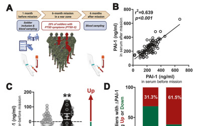
Stress-induced plasminogen activator inhibitor-1 (PAI-1) as a blood biomarker and brain risk factor for PTSD.
Mennesson M, Abdelkaoui S, Roullot-Lacarrière V, Tronel S, Cathala A, Lalanne V, Raux PL, Makrini L, Valjent E, Duffaud AM, Claverie D, Vallée M, Desmedt A, Trousselard M, Revest JM
doi: 10.1038/s41380-026-03564-w

Abstract:
Post-traumatic stress disorder (PTSD) is a severe stress-related psychiatric condition triggered by traumatic life-threatening events, characterized notably by an altered memory profile. Although clinically well-documented, no specific biomarker exists. This translational study identifies plasminogen activator inhibitor-1 (PAI-1) as a brain risk factor for PTSD, thereby supporting its potential as a blood-derived biomarker. Mice with genetically ablated PAI-1 were protected from developing a PTSD-like memory profile. Conversely, mice exhibiting PTSD-like cognitive impairment showed increased blood PAI-1 levels, correlating with their profile severity. In the brain, PAI-1 levels were specifically increased in the dorsal hippocampus, a key region for cognitive functions and in the etiology of PTSD. Finally, a longitudinal study of soldiers revealed that those developing PTSD symptoms exhibit rising blood PAI-1 levels over a 12-month period. Its significant association with various indicators of PTSD-related psychological distress attests to PAI-1's potential as a blood biomarker and brain therapeutic target for PTSD.


11/03/2026 | eur ann otorhinolaryngol head neck dis
Assessment of Micro-TRAIN simulation in otologic microsurgery.
Julian T, Bonnard D, Gimenez T, Franco-Vidal V
doi: 10.1016/j.anorl.2026.02.008

Abstract:
OBJECTIVES: Temporal bone drilling simulators are valuable tools for developing anatomical knowledge and drill handling motor skills. However, there are currently no simulators focusing on learning fine microsurgical gestures using micro-instruments. The aim of this study was to validate a training kit and simulation program dedicated to otologic microsurgery. MATERIALS AND METHODS: A prospective single-center comparative study was conducted. The Micro-TRAIN kit comprised a mannequin and six interchangeable exercise modules. Two simulation sessions were conducted two months apart, with debriefing for two selected subgroups after the first session. Progression in performance, assessed by task completion time, execution time and technical skills, was evaluated in three groups: 10 novices, 10 intermediate level and 5 experts. Content validity, face validity and participant satisfaction were also assessed. RESULTS: In session 1, there were significant differences in performance between the three groups (P<0.005). Both the novice and intermediate groups showed improvement between sessions. Improvement in total score in the intermediate group was statistically significant (P=0.0019) and suggestive in the novice group (P=0.0059). Participants who received debriefing tended to improve more (novices: P=0.012; intermediate: P=0.036). Experts rated the simulator's realism and relevance as above 8/10. CONCLUSION: This study confirmed the content, face and construct validity of the Micro-TRAIN simulator. It is an effective tool for acquiring microsurgical skills in otologic surgery.


03/2026 | fr j urol
Risk factors for RNA integrity in fresh-frozen renal cancer samples from routine practice (UroCCR 9).
Margue G, Capon G, Niort BC, Donon L, Soulet F, Quemener C, Merlio JP, Letenneur L, Doussau A, Jullien de Pommerol M, Drutel G, Gross-Goupil M, Yacoub M, Bikfalvi A, Bernhard JC

Abstract:
INTRODUCTION: High-quality biospecimens are essential for transcriptomic analyses in renal cell carcinoma (RCC), yet real-world determinants of RNA integrity in routine surgical settings remain insufficiently documented. This study aimed to evaluate RNA quantity and quality from cryopreserved RCC tissues and identify factors associated with RNA degradation. MATERIAL AND METHODS: We conducted a retrospective analysis of 160 patients who underwent surgery for clear cell RCC and included in the UroCCR network (2005-2012). Tumor and matched normal kidney tissues were prospectively collected during partial or radical nephrectomy and snap-frozen. RNA was extracted using an automated protocol. RNA concentration was measured by NanoDrop spectrophotometry, and RNA quality was assessed using the RNA Quality Score (RQS). Multivariate linear regression models with random intercepts were used to identify factors associated with RNA degradation. RESULTS: RNA extraction was successful in 302 of 310 samples. RNA concentration exceeded 25ng/muL in 93% of samples, and 78.4% met both quantitative and qualitative criteria (RQS>/=6). Mean RNA concentration was 128.8+/-104.4ng/muL, and mean RQS was 7.52+/-1.40. Tumor tissue showed higher RNA quality than matched healthy tissue (mean RQS: 8.33+/-1.02 vs. 6.66+/-1.30; P<0.001). Preoperative embolization was independently associated with lower RNA quality (P<0.001). Freezing delay (mean 30+/-25min) and duration of storage were not associated with RNA degradation. CONCLUSION: High-quality RNA can be extracted from fresh-frozen RCC tissues under routine clinical conditions. RNA integrity is mainly influenced by tumour biology rather than procedural delays, supporting biologically informed biobanking strategies.


25/02/2026 | alzheimers res ther
APOE-epsilon4 genotype and western diet synergistically aggravate synaptic dysfunction in Alzheimer's disease via D-serine disruption.
Matos M, Oliveira A, Matias I, Le Boulch A, Ciofi P, Dupuy L, Huc E, Oliet SHR, Panatier A
doi: 10.1186/s13195-026-01992-y


29/01/2026 | j neuroinflammation
Astrocyte CB(1) receptors drive blood-brain barrier disruption in central nervous system inflammatory disease.
Colomer T, Bernal-Chico A, Sanchez-Martin E, Moreno-Garcia A, Baraibar AM, Uribe-Irusta A, Iriarte-Sarria A, Beriain S, Skupio U, Gatuingt-Chasseriaud C, Gonzales D, Laplagne G, Serrat R, de Guevara IP, Matute C, Clemente D, Tepavcevic V, Fernandez-Moncada I, Chapouly C, Marsicano G, Mato S
doi: 10.1186/s12974-026-03708-3


01/2026 | j extracell biol
Placental Extracellular Vesicles Exhibit Reduced Neurogenic Potential Linked to Changes in Their miRNA Landscape Upon HCMV Infection.
Martin C, Martin H, Bergamelli M, Lobjois L, Franco L, Bordes E, Benchoua A, Balor S, Kantar D, Coyaud E, Martins F, Favereaux A, Malnou CE
doi: 10.1002/jex2.70108

Abstract:
Extracellular vesicles (EVs) are key mediators of maternal-foetal communication, regulating placental function and foetal development through the transfer of bioactive molecules. Although placental EVs play a crucial role in placental function during pregnancy, their contribution to foetal development, notably foetal brain, remains poorly understood. Human cytomegalovirus (HCMV) is the most common virus transmitted in utero and a leading cause of infectious brain malformations. Although certain central nervous system lesions caused by HCMV are explained, the neuropathogenesis of congenital infection remains poorly understood. In this study, we demonstrate that EVs from healthy placentas promote neurogenesis. However, EVs from HCMV-infected placentas lose this neurogenic potential, impairing differentiation and migration of neural stem cells, perturbations that may contribute to the neurodevelopmental defects observed in congenital HCMV infections. miRNA profiling revealed profound infection-induced changes, including the incorporation of viral miRNAs and dysregulation of host miRNAs involved in neurogenesis. These findings highlight the critical role of placental EVs in foetal brain development and their contribution to HCMV neuropathogenesis.


2026 | genet med open
Methodology of DNA extraction and sequencing from living cardiomyocytes collected by catheter in humans.
Ader F, Guilbeau-Frugier C, Lhuillier E, Martins F, Gonzalez AA, Rollin A, Beneyto M, Gales C, Pires F, Maoret JJ, Sénard JM, Timnou-Bekouti J, Villard E, Duboscq-Bidot L, Gandjbakhch E, Maury P
doi: 10.1016/j.gimo.2025.103473

Abstract:
PURPOSE: We present here the technical feasibility of percutaneously retrieving cardiomyocytes (CMs) through the lumen of irrigated ablation catheters, with the aim of obtaining DNA of sufficient quality/quantity for allowing DNA amplification, screening, and derived genetic analysis. METHODS: Irrigated conventional catheters for ablation were used for creating endocardial right ventricular voltage maps in 38 patients with suspected or proved arrhythmogenic right ventricular cardiomyopathy. Blood material was collected from scar areas by aspiration and filtered, CMs detected by light microscopy were aspirated, centrifugated, and freezed.DNA was extracted, amplified, and sequenced, and variants were compared with variants obtained from leukocyte DNA. RESULTS: At least 1 CM was obtained in 95% of patients (median 11 CM/patient). After refinements of the technique, a total of 136 samples (22 patients) allowed DNA extraction and amplification, successful in 60% of samples (16 patients). DNA capture sequencing of a panel of cardiomyopathy-associated genes was successfully performed in 14 patients and compared with blood sequencing in 11. After controlled by Sanger, an additional variant, not present in blood, has been confirmed in CM in one patient. CONCLUSION: This new mini-invasive technique of sampling allows to perform genetic analysis on CMs. Pending future improvements, this technique could provide new sources of human cells for research and potential mosaicism detection.


05/12/2025 | Nat Commun
Early intrinsic excitability plasticity of neocortical engram neurons defines memory formation and precision.
Hadzibegovic S, Zhu L, Ginger M, Gueidao Costa M, Alvarez Menendez P, De Sa R, Le Corf K, Le Feuvre Y, Nicole O, Bontempi B, Frick A
doi: 10.1038/s41467-025-66975-3

Abstract:
Neocortical memory engrams are thought to mature via strengthened interconnectivity, yet synaptic plasticity alone cannot explain the dynamic vividness of enduring memories. Neuronal intrinsic excitability (IE) plasticity has been touted as an early priming mechanism that renders engram neurons susceptible to ongoing plastic processes and later encoding events. Here, we reveal that learning-induced IE plasticity of nascent anterior cingulate cortex (ACC) engram neurons is a permissive mechanism for the formation and specificity of remote memories. Using c-fos-dependent genetic/viral targeting in mice, we found that contextual fear learning triggered a time-limited increase in ACC engram IE during the early phase of memory formation. Remarkably, chemogenetically hyperpolarizing these neurons within-but not outside-the IE plasticity window strengthened consolidated memories, enhanced their context-precision, and prevented interference-induced engram reallocation. Thus, IE plasticity in nascent ACC engram neurons acts as an essential tagging mechanism that determines the fate and dynamic content of remote memories.


24/10/2025 | adv sci (weinh)
Nanoscopic Mapping of the Extracellular Space in Amyloid Plaque-rich Cortex.
Estaún-Panzano J, Dembitskaya Y, Calaresu I, Nandi S, Gresil Q, Doudnikoff E, Leste-Lasserre T, Amédée T, Cognet L, Groc L, Nägerl UV, Bezard E
doi: 10.1002/advs.202515674

Abstract:
A hallmark of Alzheimer's disease (AD) is the accumulation of amyloid plaques, primarily composed of misfolded amyloid β (Aβ) peptides. Complementary high-resolution imaging techniques are employed to investigate the plaque penetrability and the extracellular space (ECS) rheology in a mouse model of AD. Two-photon shadow imaging in vivo confirms that a dense ring of cells surrounds cortical amyloid plaques but highlights the diffusional penetrability of the amyloid core. Quantum dot tracking unveils that ECS diffusional parameters are heterogeneous in and around plaques, with an elevated diffusivity within and around plaques compared to wild-type-tissue. The amyloid core shows low nanoparticle density, varying by plaque phenotype. Carbon nanotube tracking confirms these altered local rheological properties at the level of the whole cortex of AD mice. Finally, the extracellular matrix is found to be dysregulated within the amyloid plaque, which may account for the observed alterations in diffusivity. This study provides fresh insights for understanding Aβ plaque penetration, a prerequisite for therapeutic development.


14/10/2025 | neurochem res
D-Serine's Journey Between Stars and Synapses.
Mountadem S, Oliet SHR, Panatier A
doi: 10.1007/s11064-025-04564-y

Abstract:
Astrocytes play a pivotal role in regulating synaptic transmission, with D-serine emerging as a key gliotransmitter shaping NMDA receptor-dependent functions. This review is focusing on the multifaceted role of astrocytic D-serine from synaptic transmission to cognitive processes. While this review includes the work of other groups, it is mainly based on the findings obtained in our laboratory. Drawing from two decades of research spanning from the hypothalamus to the hippocampus, we here highlight how astrocyte-derived D-serine regulates NMDAR activity, long-term synaptic plasticity, and associated memory. Our findings have revealed the dynamic control exerted by astrocytic processes onto D-serine availability within the synaptic cleft, including the impact of the astrocytic morphological plasticity, the key role played by intracellular Ca(2+) as well as the involvement of CB1 and EphB3 receptors. We also discuss how an impairment in astrocytic D-serine synthesis can affect the co-agonist availability and consequently impact cognitive functions in neurodegenerative disorders such as Alzheimer's Disease. To conclude, this review highlights the role of astrocytic D-serine in astrocyte-neuron communication and higher-order brain functions.


01/10/2025 | Mol Psychiatry
Long-lived adult-born hippocampal neurons promote successful cognitive aging.
Blin N, Charrier V, Farrugia F, Palhol J, Presset A, Cartier E, Oliet S, Pacary E, Koehl M, Lie DC, Masachs N, Abrous DN

Abstract:
Aging is commonly associated with a decline in memory abilities, yet some individuals remain resilient to such changes. Memory processing has been shown to rely on adult neurogenesis, a form of hippocampal plasticity, but whether the integration and role of long-lived adult-born neurons (ABNs) generated during early adult life also contribute to cognitive resilience and to such inter-individual differences remain unknown. Using a pseudo-longitudinal approach in rats characterized as resilient or vulnerable to cognitive aging, we examined the survival, senescence, morphology, glutamatergic connectivity, and mitochondrial health of ABNs. To achieve this, we combined approaches based on thymidine analogues and retroviral labeling using Moloney murine leukemia viruses. While ABNs survival, entry into senescence and dendritic gross morphology did not differ between resilient and vulnerable rats, resilient animals exhibited preserved glutamatergic synaptic input and maintained mitochondrial homeostasis in the proximal dendrites of ABNs. Interestingly, bypassing this reduction in glutamatergic inputs in vulnerable rats through direct optogenetic stimulation was sufficient to rescue their memory retrieval abilities, indicating that ABNs themselves remain intrinsically functional despite reduced input. Overall, our data indicate that maintaining long-lived ABNs within the neuronal network is essential for successful cognitive aging, highlighting their potential as a therapeutic target for restoring cognitive functions in old age.


29/09/2025 | neural regen res
Astrocytic ion channel Kir4.1 deficit underlies chronic pain.
Mountadem S, Voisin DL, Dallel R
doi: 10.4103/NRR.NRR-D-25-00773


19/09/2025 | rev endocr metab disord
Beyond satiety: unraveling the complex roles of POMC neurons in behavior and metabolism.
Jouque V, Miralpeix C, Lopez-Gambero AJ, Nicolas JC, Quarta C, Cota D
doi: 10.1007/s11154-025-09993-2

Abstract:
Hypothalamic pro-opiomelanocortin (POMC) neurons are classically viewed as mediators of satiety, acting in response to metabolic and hormonal cues and in opposition to Agouti-related protein (AgRP) neurons to maintain energy balance. This model, centered on the appetite-suppressant effects of the POMC-derived neuropeptide alpha-melanocyte-stimulating hormone (alpha-MSH) through its activation of melanocortin-4 receptors (MC4R), has shaped our understanding of feeding and body weight regulation for decades. However, recent discoveries have challenged and expanded this traditional view, revealing that POMC neurons are not a uniform population dedicated solely to satiety control. Single-cell transcriptomic analyses have revealed striking molecular heterogeneity, reflected in distinct anatomical distributions, receptor expression profiles, electrophysiological properties, and projection patterns - all supporting the idea of functional specialization within this neuronal population. In this review, we propose a conceptual framework that integrates POMC neuronal heterogeneity with the regulation of appetite, metabolic physiology, and behavior beyond feeding. We highlight emerging evidence showing that discrete POMC neuronal subpopulations respond to specific combinations of interoceptive and environmental cues to orchestrate diverse adaptive responses. This perspective underscores the developmental plasticity and functional versatility of POMC neurons, offering new insights into the mechanisms of obesity and potentially paving the way for novel targeted therapeutic strategies.


19/09/2025 | Eur J Endocrinol
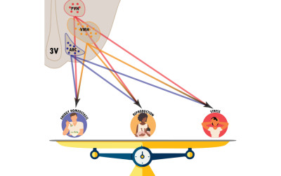
New routes to the neuroendocrine hypothalamus: the extracellular space.
Nicolas JC, Huwart SJP, Ziemens D, Freire-Agulleiro O, Lee TH, Mattot V, Quarta C
doi: 10.1093/ejendo/lvaf197

Abstract:
The neuroendocrine hypothalamus integrates peripheral nutritional and hormonal cues to regulate essential physiological processes, including appetite, metabolism and reproduction. While the mechanisms by which hormones traverse the blood-brain barrier to access the hypothalamic parenchyma are well characterised, how these signals subsequently diffuse and distribute within the brain's extracellular space and matrix remains poorly understood. Emerging evidence implicates specialised components of the extracellular matrix, such as perineuronal nets (PNNs), in modulating hormonal and nutrient bioavailability, as well as neuronal excitability and plasticity. In the hypothalamus, extracellular matrix components are highly dynamic and respond to nutritional and hormonal cues. In preclinical models of metabolic disorders involving the neuroendocrine system - such as obesity and type 2 diabetes - these components undergo maladaptive remodelling. This Review discusses recent advances in our understanding of how the extracellular environment shapes neuroendocrine signalling in the hypothalamus, and explores the broader implications for systemic hormonal regulation and neuroendocrine disease pathophysiology.


09/2025 | Nat Neurosci
Potentiation of mitochondrial function by mitoDREADD-G(s) reverses pharmacological and neurodegenerative cognitive impairment in mice.
Pagano Zottola AC, Martin-Jimenez R, Lavanco G, Hamel-Cote G, Ramon-Duaso C, Rodrigues RS, Mariani Y, Khan M, Drago F, Jean S, Rio IB, Jimenez-Blasco D, Egana-Huguet J, Eraso-Pichot A, Beriain S, Cannich A, Vidal-Palencia L, Infantino R, Julio-Kalajzic F, Gisquet D, Goncalves A, Al-Younis I, Baussan Y, Duvezin-Caubet S, Devin A, Soria-Gomez E, Puente N, Bolanos JP, Grandes P, Pouvreau S, Busquets-Garcia A, Marsicano G, Bellocchio L, Hebert-Chatelain E

Abstract:
Many brain disorders involve mitochondrial alterations, but owing to the lack of suitable tools, the causal role of mitochondrial dysfunction in pathophysiological processes is difficult to establish. Heterotrimeric guanine nucleotide-binding (G) proteins are key regulators of cell functions, and they can be found within mitochondria. Therefore, we reasoned that the activation of stimulatory mitochondrial G proteins (G(s)) could rapidly promote the activity of the organelle and possibly compensate for bioenergetic dysfunction. Here, we show that a mitochondria-targeted recombinant designer receptor exclusively activated by designer drugs (mitoDREADD-G(s)) can acutely trigger intramitochondrial signaling to increase mitochondrial membrane potential and oxygen consumption. In vivo activation of mitoDREADD-G(s) abolished memory alterations in cannabinoid-treated mice and in two mouse models of Alzheimer's disease and frontotemporal dementia. Thus, mitoDREADD-G(s) enables the establishment of causal relationships between mitochondria and biological or disease-related processes and represents an innovative potential therapeutic approach for disorders associated with mitochondrial impairment.


Abstract:
While blood vessels and osteocytes have been studied independently, their simultaneous changes with age remain undescribed. Our objective was to investigate the age-related evolution of both osteocyte and blood vessel networks in mouse cortical bone, and to assess the associated effects on osteocyte markers and oxygen intracellular levels. We analyzed femurs of male Flk1-GFP mice from growing, mature, middle-aged, and aged groups with techniques such as laser microdissection followed by RT-qPCR, tissue clearing and 3D fluorescence imaging. In the mature animals - when the cortical bone was thicker than in the growing animals - the osteocyte density, the number of dendrites per osteocyte and the blood vessel density were lower. This was associated with a reduced expression of Pdpn and with a smaller fraction of osteocytes exhibiting low intracellular oxygen. In aged animals - when cortical bone was thinner than in mature animals - the number of dendrites per osteocyte and the blood vessel density were lower. This was associated with a reduced Gja1 (Cx43) expression. Our results suggest that changes in the osteocyte network during maturation and aging are led by distinct mechanisms, and that the cortical bone blood vessels are not the main source of oxygen for osteocytes.


07/2025 | neurobiol stress
Chronic stress and cytogenesis ablation disrupt hippocampal neuron connectivity, with fluoxetine restoring function with sex-specific effects.
Ribeiro I, Silveira-Rosa T, Martins-Macedo J, Marques-Ferraz L, Dourado AR, Martins-Ferreira G, Farrugia F, Rodrigues AJ, Abrous DN, Alves ND, Patricio P, Pinto L

Abstract:
Hippocampal Adult-Born neurons (hABNs) play a critical role in maintaining brain homeostasis, exhibiting unique properties during their maturation. The absence of hABNs impacts surrounding neuronal networks, but the mechanisms are not fully understood. In this study, we examined how perturbations to adult hippocampal cytogenesis affect the neuronal inputs to adult-born and mature neurons in the dentate gyrus. In particular, we analyzed connectivity changes in GFAP-Tk transgenic rats with ablated neurogenesis, and in unpredictable Chronic Mild Stress (uCMS) rats with reduced neurogenesis, with a focus on sex-differences in stress-affected hABNs. Additionally, we evaluated the action of the antidepressant fluoxetine on brain connectivity. Using a virus-mediated retrograde tracing approach, we quantified synaptic inputs to mature neurons and hABNs. Our findings reveal significant impairments in both intra- and extra-hippocampal connectivity following disruptions in cytogenesis, providing new insights into hippocampal network dynamics in the context of cytogenetic impairment, depression, and therapeutic interventions.


24/06/2025 | neurochem res
The Endocannabinoid System in Retinal Muller Glia: Lessons From Astrocyte Research.
Beriain S, Fernandez-Moncada I, Pereiro X, Eraso-Pichot A, Vecino E, Marsicano G
doi: 10.1007/s11064-025-04457-0

Abstract:
The endocannabinoid system (ECS) is a widespread neuromodulatory system involved in both physiological and pathological processes. The ECS has been mainly characterized in neurons, but emerging evidence has revealed its presence in different glial cells. Many studies focused on the functions of the ECS in astrocytes, but its role in other glial cells remains poorly explored. Muller cells are the main glial cells in the retina, providing structural maintenance, metabolic support and neuroprotection to other retinal cells. However, the crosstalk between the ECS and Muller glia (MG) has been scantly described so far. In this context, inspired by similar research on astrocytes, the study of the presence and activity of the ECS in MG is currently emerging as an interesting way of controlling retinal functions in physiopathological conditions. This review will examine the current knowledge on the ECS in MG with the general aim to promote future research in this field.


20/06/2025 | iscience
Bronchial smooth muscle extracellular vesicles interfere with bronchial epithelium metabolism and function in asthma.
Celle E, Chahin A, Beaufils F, Cardouat G, Eyraud E, Bouchet C, Campagnac M, Ousova O, Begueret H, Thumerel M, Dubois R, Dupuy JW, Leste-Lasserre T, Lacomme S, Lager-Lachaud N, Bellvert F, Marthan R, Girodet PO, Berger P, Trian T, Esteves P
doi: 10.1016/j.isci.2025.112546

Abstract:
Bronchial smooth muscle (BSM) remodeling is an important feature of severe asthma pathophysiology. We previously showed that asthmatic BSM is metabolically different and increased rhinovirus (RV) replication rate, the main trigger of severe asthma exacerbations. Extracellular vesicles (EVs) are the key mediator in cell-cell communication, but the role of BSM cells-derived EVs on bronchial epithelial has never been investigated in asthma. Using severe asthmatic and non-asthmatic tissue collection, we show that asthmatic BSM cells are able to produce a greater amount of EVs containing metabolites involved in bioenergetics. We study the bronchial epithelium energetic rewiring following stimulation with asthmatic BSM cells-derived EVs. Modifications of bronchial epithelium metabolic behavior were associated with an increased ATP production and a breakdown of bronchial epithelium barrier function such as ciliary beating frequency and efficiency. Finally, we show that asthmatic BSM cells-derived EVs increased RV replication in bronchial epithelium following RV infection.


12/06/2025 | J Clin Invest
MuSK cysteine-rich domain antibodies are pathogenic in a mouse model of autoimmune myasthenia gravis.
Halliez M, Cottin S, You A, Buon C, Grondin A, Lippens LS, Lemaitre M, Ezan J, Isch C, Rufin Y, Montcouquiol M, Sans N, Fontaine B, Messéant J, Le Panse R, Strochlic L
doi: 10.1172/JCI173308

Abstract:
The neuromuscular junction (NMJ), synapse between the motor neuron terminal and a skeletal muscle fiber is crucial, throughout life, in maintaining the reliable neurotransmission required for functional motricity. Disruption of this system leads to neuromuscular disorders, such as auto-immune myasthenia gravis (MG), the most common form of NMJ diseases. MG is caused by autoantibodies directed mostly against the acetylcholine receptor (AChR) or the muscle-specific kinase MuSK. Several studies report immunoreactivity to the Frizzled-like cysteine-rich Wnt-binding domain of MuSK (CRD) in patients, although the pathogenicity of the antibodies involved remains unknown. We showed here that the immunoreactivity to MuSK CRD induced by the passive transfer of anti-MuSKCRD antibodies in mice led to typical MG symptoms, characterized by a loss of body weight and a locomotor deficit. The functional and morphological integrity of the NMJ was compromised with a progressive decay of neurotransmission and disruption of the structure of pre- and post-synaptic compartments. We found that anti-MuSKCRD antibodies completely abolished Agrin-mediated AChR clustering by decreasing the Lrp4-MuSK interaction. These results provide the first demonstration of the role of the MuSK CRD in MG pathogenesis and improve our understanding of the underlying pathophysiological mechanisms.


06/2025 | Mol Metab
CPT1C deficiency in SF1 neurons impairs early metabolic adaptation to dietary fats, leading to obesity.
Fosch A, Pizarro DS, Zagmutt S, Reguera AC, Batallé G, Rodríguez-García M, García-Chica J, Freire-Agulleiro O, Miralpeix C, Zizzari P, Serra D, Herrero L, López M, Cota D, Rodríguez-Rodríguez R, Casals N

Abstract:
OBJECTIVES: SF1 neurons of the ventromedial hypothalamus (VMH) play a pivotal role in regulating body weight and adiposity, particularly in response to a high-fat diet (HFD), as well as in the recovery from insulin-induced hypoglycemia. While the brain-specific CPT1C isoform is well known for its role in controlling food intake and energy homeostasis, its function within specific hypothalamic neuronal populations remains largely unexplored. Here, we explore the role of CPT1C in SF1 neurons. METHODS: Mice deficient in CPT1C within SF1 neurons were generated, and their response to a HFD was investigated. RESULTS: SF1-Cpt1c-KO mice fail to adjust their caloric intake during initial HFD exposure, which is associated with impaired activation of the melanocortin system. Furthermore, these mice exhibit disrupted metabolic gene expression in the liver, muscle, and adipose tissue, leading to increased adiposity independently of food intake. In contrast, their response to glucose or insulin challenges remains intact. After long-term HFD exposure, SF1-Cpt1c-KO mice are more prone to developing obesity and glucose intolerance than control littermates, with males exhibiting a more severe phenotype. Interestingly, CPT1C deficiency in SF1 neurons also results in elevated hypothalamic endocannabinoid (eCB) levels under both chow and HFD conditions. We propose that this sustained eCB elevation reduces VMH activation by fatty acids and impairs the SF1-POMC drive upon fat intake. CONCLUSION: Our findings establish CPT1C in SF1 neurons as essential for VMH-driven dietary fat sensing, satiety, and lipid metabolic adaptation.


26/05/2025 | Mol Neurobiol
Nociceptin/OrphaninFQ Receptor Modulates the Maturation of Adult-Born Neurons in the Mouse Dentate Gyrus Under Physiological Conditions and in a Chronic Stress Model.
Robert C, D'Oliveira da Silva F, Seminara F, Martinelli C, Farrugia F, Sturaro C, Pacary E, Rampon C, Ruzza C, Mouledous L

Abstract:
Neurogenesis persists in the adult dentate gyrus (DG) of the hippocampus, playing a critical role in memory and stress adaptation. Dysregulation of this process is implicated in cognitive deficits and depressive behaviors induced by chronic stress, while classical antidepressants are known to enhance neurogenesis. The Nociceptin/Orphanin FQ (N/OFQ) system, comprising N/OFQ and its NOP receptor, modulates memory and the stress response, yet its role in adult neurogenesis remains underexplored. Here, we investigated the impact of N/OFQ signaling on neurogenesis in the mouse DG using genetic and pharmacological approaches under basal and chronic stress conditions. In constitutive NOP receptor knockout (KO) mice, adult neurogenesis was only mildly altered, with subtle changes in neuronal maturation. However, spine density in 4-week-old adult-born DG neurons increased following conditional NOP Receptor KO in the DG. The increase was specific to stubby and thin spines, while mature mushroom spine density decreased. When NOP KO was restricted to newly born neurons, no significant differences were observed in spine density suggesting that the absence of NOP receptors in mature DG neurons influences the local environment to regulate spinogenesis in adult-born neurons indirectly. Finally, chronic corticosterone exposure impaired spinogenesis in immature neurons, and this was mitigated by systemic administration of a NOP antagonist. Our findings suggest that N/OFQ signaling indirectly regulates the maturation and connectivity of adult-born neurons through modulation of local and distal inputs. This regulation may contribute to the antidepressant and pro-cognitive effects of NOP receptor antagonists.


28/04/2025 | Glia
Astroglial CB(1) Reveal Sex-Specific Synaptic Effects of Amphetamine.
Mariani Y, Dalla-Tor T, Garavaldi T, Julio-Kalajzic F, Gisquet D, Gomez-Sotres P, Cannich A, Gambino G, Drago F, Serrat R, Hurel I, Chaouloff F, Pouvreau S, Bellocchio L, Marsicano G, Covelo A
doi: 10.1002/glia.70026

Abstract:
The Nucleus Accumbens (NAc) is a critical brain region for the effects of psychostimulant drugs. Type-1 cannabinoid receptors (CB(1)), the main elements of the endocannabinoid system (ECS) in the brain, participate in these effects and modulate synaptic functions in the NAc. Besides their neuronal expression, CB(1) receptors are also present in astrocytes, where they contribute to the regulation of synaptic plasticity and behavior. However, the impact of astroglial CB(1) receptors on synaptic plasticity in the NAc and on psychostimulant-induced synaptic and behavioral effects is currently unknown. This study shows that the psychostimulant amphetamine impairs a form of astroglial CB(1) receptor-dependent synaptic plasticity in the NAc of male, but not female mice. Consistently, locomotor effects of amphetamine require astroglial CB(1) receptors in male, but not female mice. These results, by revealing unforeseen mechanisms underlying sex-dependent effects of amphetamine, pave the way to a better understanding of the diverse impact of psychostimulants in women and men.


11/04/2025 | Diabetes
GLP-1-mediated targeting of inflammation corrects obesogenic memory in male mice.
Leon S, Benoit J, Clark S, Zizzari P, Yang B, Dugail I, Merabtene F, Clement K, Eygret L, Dupuy N, Delpech JC, Rossitto M, Mack M, Leste-Lasserre T, Finan B, Cota D, Quarta C
doi: 10.2337/db24-1071

Abstract:
Obesity-induced biological changes often persist after weight loss and are difficult to reverse, a phenomenon known as 'obesogenic memory'. This enduring effect is associated with metabolic inflammation, particularly in adipose tissue. In this study, we characterise a mouse model of obesogenic memory and evaluate the efficacy of the unimolecular conjugate GLP-1/Dexa, which selectively and safely delivers the anti-inflammatory drug dexamethasone to GLP-1 receptor (GLP-1R)-expressing cells. We document that this precision pharmacological approach outperforms treatment with GLP-1 or dexamethasone alone, significantly reducing body weight, food intake, adiposity and markers of adipose tissue inflammation in male mice with obesogenic memory. In addition, we identify the CCR2/CCL2 inflammatory pathway as an important mediator of glucose intolerance and adipose tissue inflammation associated with obesogenic memory. Our findings suggest that targeting inflammation via GLP-1R signalling may be a promising therapeutic strategy to alleviate obesogenic memory and improve the long-term clinical management of metabolic diseases.


04/04/2025 | Brain
Astrocytic Kir4.1 ion channel deficit drives persistent inflammatory facial pain in males.
Mountadem S, Herault K, Peirs C, da Silva Borges G, Voisin DL, Antri M, Dallel R
doi: 10.1093/brain/awaf122

Abstract:
Chronic facial pain, a frequent and disabling condition, is maintained by central sensitization, which results in pain hypersensitivity. Although it is well established that reactive astrocytes play a key role in persistent pain mechanisms, the role of disruption of the normal capacity of astrocytes to maintain neuronal homeostasis is much less known. Here we show that persistent facial inflammation disturbs potassium homeostasis in the medullary dorsal horn of male rats, due to a sex-specific, drastic downregulation of astroglial inward rectifier potassium Kir4.1 channels. Using selective genetic tools, we establish that such downregulation, likely due to the release of IL-1beta during inflammatory processing, is sufficient and required to drive pain hypersensitivity through altered K+ baseline levels. We further show that this chain of events can be prevented by selective upregulation of astroglial Kir4.1, or through systemic administration of 5-azacytidine, a DNA methylation modulator. Our results thus reveal a critical mechanism by which astrocyte dysfunction drives persistent inflammatory facial pain in males and discover the therapeutic potential of targeting central Kir4.1 for treating this disease.


14/03/2025 | Neurobiol Dis
Developmental alterations of indirect-pathway medium spiny neurons in mouse models of Huntington's disease.
Lebouc M, Bonamy L, Dhellemmes T, Scharnholz J, Richard Q, Courtand G, Brochard A, Martins F, Landry M, Baufreton J, Garret M
doi: 10.1016/j.nbd.2025.106874

Abstract:
Huntington's disease (HD) is a complex neurodegenerative disorder with cognitive and motor symptoms that typically manifest in adulthood. However, embryonic brain development impairments leading to cortical defects in HD mutation carriers has been shown recently supporting a neurodevelopmental component in HD. Despite HD is primarily recognized as a striatal pathology, developmental alterations in this structure, particularly during the early postnatal period, remain poorly understood. To fill this gap, we examined striatal development in newborn R6/1 mice. We found that D2 receptor-expressing indirect-pathway medium spiny neurons (D2-MSNs) present in the matrix striatal compartment undergo early morphological and electrophysiological maturation. Altered electrophysiological properties were also observed in newborn CAG140 mice. Additionally, we also observed a D2-MSN-selective reduction in glutamatergic cortico-striatal transmission at the beginning of the second postnatal week as well as a reduced projection of D2-MSNs onto the GPe at birth in R6/1 mice. All these alterations were transient with the circuit normalizing after the second postnatal week. These results identify a compartment- and cell-type specific defect in D2-MSNs maturation, which can contribute in their latter vulnerability, as this cell-type is the first to degenerate in HD during adulthood.


12/03/2025 | Brain
MRI R2* captures inflammation in disconnected brain structures after stroke: a translational study.
Koubiyr I, Yamamoto T, Petit L, Dubourdieu N, Avignone E, Cozensa E, Galmiche C, Fukutomi H, Sibon I, Dousset V, Thiebaut de Schotten M, Panatier A, Tible M, Tourdias T
doi: 10.1093/brain/awaf082

Abstract:
Ischemic strokes disrupt brain networks, leading to remote effects in key regions like the thalamus, a critical hub for brain functions. However, non-invasive methods to quantify these remote consequences still need to be explored. This study aimed to demonstrate that MRI-derived R2* changes can capture iron accumulation linked with inflammation secondary to stroke-induced disconnection. In order to link remote R2* changes to stroke-induced disconnection, we first conducted a secondary analysis of 156 prospectively included stroke patients who underwent MRI at baseline and 1-year follow-up. We mapped fibers disconnected by baseline infarcts to compare the R2* changes over 1 year according to the disconnectivity status in specific thalamic nuclei groups. We also identified the variables associated with elevated R2* at 1 year in a multivariate context through linear regressions. In parallel, to understand the biological underpinning of the remote R2* changes, we set up a translational mouse model through photothrombotic induction of focal cortical infarcts or sham procedures in 110 C57BL/6J mice. We explored the mice through combinations of in vivo MRI at 72h, 2-, 4-, and 8-weeks, histology, qPCR for gene expression, mass spectrometry for iron concentration quantification, and additional ex vivo high-resolution diffusion tensor imaging. In stroke patients, we found a significant increase of R2* within severely disconnected medial and lateral thalamic nuclei groups from baseline to 1 year. At the same time, no change occurred if these structures were not disconnected. We also showed that the disconnectivity status at baseline was significantly associated with R2* at follow-up, independently from confounders, establishing a direct and independent relationship between baseline disconnection and the subsequent R2* increase within the associated locations. In mice, we recapitulated the patients' conditions by observing increased R2* in the stroke groups, specifically within the disconnected thalamic nuclei. Such remote and focal R2* changes peaked at 2 weeks, preceding and correlating with longer-term atrophy at 8 weeks. We established that the remote R2* increase was spatially and temporally correlated with a significant increase of chemically determined iron load bound to ferritin within activated microglial cells. This study provides critical evidence that R2* is a sensitive marker of inflammation secondary to network disconnection, potentially informing future neuroprotective strategies targeting remote brain regions after stroke.


11/03/2025 | Prog Neurobiol
Astrocytic EphB3 receptors regulate D-serine-gated synaptic plasticity and memory.
Langlais VC, Mountadem S, Benazzouz I, Amadio A, Matos M, Jourdes A, Cannich A, Julio-Kalajzic F, Belluomo I, Matias I, Maitre M, Leste-Lasserre T, Marais S, Avignone E, Marsicano G, Bellocchio L, Oliet SHR, Panatier A

Abstract:
The activation of classical NMDA receptors (NMDARs) requires the binding of a co-agonist in addition to glutamate. Whereas astrocytic-derived d-serine was shown to play such a role at CA3-CA1 hippocampal synapses, the exact mechanism by which neurons interact with neighboring astrocytes to regulate synaptic d-serine availability remains to be fully elucidated. Considering the close anatomical apposition of astrocytic and neuronal elements at synapses, the aforementioned process is likely to involve cells adhesion molecules. One very likely candidate could be the astrocytic EphB3 receptor and its neuronal partner, ephrinB3. Here, we first showed in acute hippocampal slices from adult mice that stimulation of EphB3 receptors with exogenous ephrinB3 increased d-serine availability at CA3-CA1 synapses, resulting in an increased NMDAR activity. Conversely, inhibiting endogenous EphB3 receptors caused an impairment of both synaptic NMDAR activity and NMDAR-dependent long-term synaptic potentiation (LTP), effects that could be rescued by exogenous d-serine. Most interestingly, knocking down EphB3 receptor specifically in astrocytes yielded a similar impairment in hippocampal plasticity and, most importantly, caused a deficit in novel object recognition memory. Altogether, our data thus indicate that EphB3 receptors in hippocampal astrocytes play a key role in regulating synaptic NMDAR function, activity-dependent plasticity and memory.


15/02/2025 | Prog Neurobiol
The correct connectivity of the DG-CA3 circuits involved in declarative memory processes depends on Vangl2-dependent planar cell polarity signaling.
Depret N, Gleizes M, Moreau MM, Poirault-Chassac S, Quiedeville A, Carvalho SDS, Venugopal V, Abed ASA, Ezan J, Barthet G, Mulle C, Desmedt A, Marighetto A, Racca C, Montcouquiol M, Sans N

Abstract:
In the hippocampus, dentate gyrus granule cells connect to CA3 pyramidal cells via their axons, the mossy fibers (Mf). The synaptic terminals of Mfs (Mf boutons, MfBs) form large and complex synapses with thorny excrescences (TE) on the proximal dendrites of CA3 pyramidal cells (PCs). MfB/TE synapses have distinctive 'detonator' properties due to low initial release probability and large presynaptic facilitation. The molecular mechanisms shaping the morpho-functional properties of MfB/TE synapses are still poorly understood, though alterations in their morphology are associated with Down syndrome, intellectual disabilities, and Alzheimer's disease. Here, we identify the core PCP gene Vangl2 as essential to the morphogenesis and function of MfB/TE synapses. Vangl2 colocalises with the presynaptic heparan sulfate proteoglycan glypican 4 (GPC4) to stabilise the postsynaptic orphan receptor GPR158. Embryonic loss of Vangl2 disrupts the morphology of MfBs and TEs, impairs ultrastructural and molecular organisation, resulting in defective synaptic transmission and plasticity. In adult, the early loss of Vangl2 results in a number of hippocampus-dependent memory deficits including characteristic flexibility of declarative memory, organisation and retention of working / everyday-like memory. These deficits also lead to abnormal generalisation of memories to salient cues and diminished ability to form detailed contextual memories. Together, these results establish Vangl2 as a key regulator of DG-CA3 connectivity and functions.


28/01/2025 | Cell Rep
Secondary motor cortex tracks decision value during the learning of a non-instructed task.
Augusto E, Kouskoff V, Chenouard N, Giraudet M, Peltier L, de Miranda A, Louis A, Alonso L, Gambino F
doi: 10.1016/j.celrep.2024.115152

Abstract:
Optimal decision-making depends on interconnected frontal brain regions, enabling animals to adapt decisions based on internal states, experiences, and contexts. The secondary motor cortex (M2) is key in adaptive behaviors in expert rodents, particularly in encoding decision values guiding complex probabilistic tasks. However, its role in deterministic tasks during initial learning remains uncertain. Here, we describe a self-initiated deterministic task requiring mice to use their forepaws to make choices without guiding cues. Our findings reveal that spontaneous decisions follow a 'race' model between actions, which uncovers underlying decision values. We use in vivo microscopy and modeling to show that M2 neurons in male mice exhibit persistent activity-encoding decision values that predict action-selection probabilities. Optogenetic inhibition of the M2 reduces the reversal performance and alters the decision value. Additionally, updates in decision values determine the rate at which learning is reversed. These results highlight the use of decision values by the M2 to adapt choice during initial learning without instructive cues.


05/12/2024 | trends endocrinol metab
Can brain neurons change identity? Lessons from obesity.
Nicolas JC, Lee TH, Quarta C
doi: 10.1016/j.tem.2024.11.006

Abstract:
It has long been thought that the functional identity of mammalian brain neurons is programmed during development and remains stable throughout adult life; however, certain populations of neurons continue to express active regulators of neuronal identity into adulthood. Prolonged exposure to diet-induced metabolic stress induces features of neuronal identity modification in adult mice, and maladaptive changes in neuronal identity maintenance have been linked to cognitive impairment in humans suffering from neurodegenerative diseases often associated with obesity. Here we discuss how, by unraveling the neurological roots of obesity, we may solve the puzzle of whether mammalian brain neurons retain identity plasticity into adulthood, while advancing knowledge of the pathogenic mechanisms at the interface of metabolic and neurodegenerative disorders.


12/2024 | clin transl med
Dynamic conditioning of porcine kidney grafts with extracellular vesicles derived from urine progenitor cells: A proof-of-concept study.
Burdeyron P, Giraud S, Lepoittevin M, Jordan N, Brishoual S, Jacquard M, Ameteau V, Boildieu N, Lemarie E, Daniel J, Martins F, Mélis N, Coué M, Thuillier R, Leuvenink H, Pellerin L, Hauet T, Steichen C
doi: 10.1002/ctm2.70095

Abstract:
 : Among strategies to limit ischemia/reperfusion (IR) injuries in transplantation, cell therapy using stem cells to condition/repair transplanted organs appears promising. We hypothesized that using a cell therapy based on extracellular vesicles (EVs) derived from urine progenitor cells (UPCs) during hypothermic and normothermic machine perfusion can prevent IR-related kidney damage. We isolated and characterized porcine UPCs and their extracellular vesicles (EVs). Then these were used in an ex vivo porcine kidney preservation model. Kidneys were subjected to warm ischemia (32 min) and then preserved by hypothermic machine perfusion (HMP) for 24 h before 5 h of normothermic machine perfusion (NMP). Three groups were performed (n = 5-6): Group 1 (G1): HMP/vehicle + NMP/vehicle, Group 2 (G2): HMP/EVs + NMP/vehicle, Group 3 (G3): HMP/EVs + NMP/EVs. Porcine UPCs were successfully isolated from urine and fully characterized as well as their EVs which were found of expected size/phenotype. EVs injection during HMP alone, NMP alone, or both was feasible and safe and did not impact perfusion parameters. However, cell damage markers (LDH, ASAT) were decreased in G3 compared with G1, and G3 kidneys displayed a preserved tissue integrity with reduced tubular dilatation and inflammation notably. However, renal function indicators such as creatinine clearance measured for 5 h of normothermic perfusion or NGAL perfusate's level were not modified by EVs injection. Regarding perfusate analysis, metabolomic analyses and cytokine quantification showed an immunomodulation signature in G3 compared with G1 and highlighted potential metabolic targets. In vitro, EVs as well as perfusates from G3 partially recovered endothelial cell metabolic activity after hypoxia. Finally, RNA-seq performed on kidney biopsies showed different profiles between G1 and G3 with regulation of potential IR targets of EVs therapy. We showed the feasibility/efficacy of UPC-EVs for hypothermic/normothermic kidney conditioning before transplantation, paving the way for combining machine perfusion with EVs-based cell therapy for organ conditioning. HIGHLIGHTS: ·UPCs from porcine urine can be used to generate a cell therapy product based on extracellular vesicles (pUPC-EVs). ·pUPC-EVs injection during HMP and NMP decreases cell damage markers and has an immunomodulatory effect. ·pUPC-EVs-treated kidneys have distinct biochemical, metabolic, and transcriptomic profiles highlighting targets of interest. ·Our results pave the way for combining machine perfusion with EV-based cell therapy for kidney conditioning.


28/11/2024 | Int J Mol Sci
The Neuroprotective Effect of the X Protein of Orthobornavirus Bornaense Type 1 in Amyotrophic Lateral Sclerosis.
Tournezy J, Leger C, Klonjkowski B, Gonzalez-Dunia D, Szelechowski M, Garenne A, Mathis S, Chevallier S, Le Masson G
doi: 10.3390/ijms252312789

Abstract:
In amyotrophic lateral sclerosis (ALS), early mitochondrial dysfunction may contribute to progressive motor neuron loss. Remarkably, the ectopic expression of the Orthobornavirus bornaense type 1 (BoDV-1) X protein in mitochondria blocks apoptosis and protects neurons from degeneration. Therefore, this study examines the neuroprotective effects of X protein in an ALS mouse model. We first tested in vitro the effect of the X-derived peptide (PX3) on motoneurons primary cultures of SOD1(G93A) mice. The total intracellular adenosine triphosphate (ATP) content was measured after incubation of the peptide. We next tested in vivo the intramuscular injection of X protein using a canine viral vector (CAV2-X) and PX3 intranasal administrations in SOD1(G93A) mice. Disease onset and progression were assessed through rotarod performance, functional motor unit analysis via electrophysiology, and motor neuron survival by immunohistochemistry. The results showed that in vitro PX3 restored the ATP level in SOD1(G93A) motor neurons. In vivo, treated mice demonstrated better motor performance, preserved motor units, and higher motor neuron survival. Although life expectancy was not extended in this severe mouse model of motor neuron degeneration, the present findings clearly demonstrate the neuroprotective potential of X protein in a model of ALS. We are convinced that further studies may improve the therapeutic impact of X protein with optimized administration methods.


25/11/2024 | Mol Metab
TGR5 receptors in SF1-expressing neurons of the ventromedial hypothalamus regulate glucose homeostasis.
Zizzari P, Castellanos-Jankiewicz A, Yagoub S, Simon V, Clark S, Maître M, Dupuy N, Leste-Lasserre T, Gonzales D, Schoonjans K, Fénelon VS, Cota D
doi: 10.1016/j.molmet.2024.102071

Abstract:
OBJECTIVE: Steroidogenic factor-1 (SF1) neurons of the ventromedial hypothalamus play key roles in the regulation of food intake, body weight and glucose metabolism. The bile acid receptor Takeda G protein-coupled receptor 5 (TGR5) is expressed in the hypothalamus, where it determines some of the actions of bile acids on food intake and body weight through still poorly defined neuronal mechanisms. Here, we examined the role of TGR5 in SF1 neurons in the regulation of energy balance and glucose metabolism. METHODS: We used a genetic approach combined with metabolic phenotyping and molecular analyses to establish the effect of TGR5 deletion in SF1 neurons on meal pattern, body weight, body composition, energy expenditure and use of energy substrates as well as on possible changes in glucose handling and insulin sensitivity. RESULTS: Our findings reveal that TGR5 in SF1 neurons does not play a major role in the regulation of food intake or body weight under standard chow, but it is involved in the adaptive feeding response to the acute exposure to cold or to a hypercaloric, high-fat diet, without changes in energy expenditure. Notably, TGR5 in SF1 neurons hinder glucose metabolism, since deletion of the receptor improves whole-body glucose uptake through heightened insulin signaling in the hypothalamus and in the brown adipose tissue. CONCLUSIONS: TGR5 in SF1 neurons favours satiety by differently modifying the meal pattern in response to specific metabolic cues. These studies also reveal a novel key function for TGR5 in SF1 neurons in the regulation of whole-body insulin sensitivity, providing new insight into the role played by neuronal TGR5 in the regulation of metabolism.


10/10/2024 | j neuroinflammation
Astrocytic DLL4-NOTCH1 signaling pathway promotes neuroinflammation via the IL-6-STAT3 axis.
Mora P, Laisne M, Bourguignon C, Rouault P, Jaspard-Vinassa B, Maitre M, Gadeau AP, Renault MA, Horng S, Couffinhal T, Chapouly C
doi: 10.1186/s12974-024-03246-w

Abstract:
Under neuroinflammatory conditions, astrocytes acquire a reactive phenotype that drives acute inflammatory injury as well as chronic neurodegeneration. We hypothesized that astrocytic Delta-like 4 (DLL4) may interact with its receptor NOTCH1 on neighboring astrocytes to regulate astrocyte reactivity via downstream juxtacrine signaling pathways. Here we investigated the role of astrocytic DLL4 on neurovascular unit homeostasis under neuroinflammatory conditions. We probed for downstream effectors of the DLL4-NOTCH1 axis and targeted these for therapy in two models of CNS inflammatory disease. We first demonstrated that astrocytic DLL4 is upregulated during neuroinflammation, both in mice and humans, driving astrocyte reactivity and subsequent blood-brain barrier permeability and inflammatory infiltration. We then showed that the DLL4-mediated NOTCH1 signaling in astrocytes directly drives IL-6 levels, induces STAT3 phosphorylation promoting upregulation of astrocyte reactivity markers, pro-permeability factor secretion and consequent blood-brain barrier destabilization. Finally we revealed that blocking DLL4 with antibodies improves experimental autoimmune encephalomyelitis symptoms in mice, identifying a potential novel therapeutic strategy for CNS autoimmune demyelinating disease. As a general conclusion, this study demonstrates that DLL4-NOTCH1 signaling is not only a key pathway in vascular development and angiogenesis, but also in the control of astrocyte reactivity during neuroinflammation.


09/10/2024 | org biomol chem
Synthesis and biological evaluation of natural Lachnophyllum methyl ester, Lachnophyllum lactone and their synthetic analogs.
Adande K, Simalou O, Ardanuy J, Eloh K, Mehalla C, Constant P, Fabing I, Genisson Y, Ballereau S

Abstract:
(2Z)-Lachnophyllum methyl ester and (4Z)-Lachnophyllum lactone were recently identified as major components in essential oils and extracts of Conyza bonariensis from Togo. Extended biological evaluation of these acetylenic compounds was however hampered by the reduced amounts isolated. A synthetic route was designed providing access to larger quantities of these two natural products as well as to original non-natural analogs with the prospect of exploring for the first time the structure-activity relationships in this series. Using LC/MS analysis, synthetic samples allowed confirming the presence of the two previously isolated natural products in plant extracts obtained by the accelerated solvent extraction technique. The nematocidal activity of the synthesized compounds confirmed the potency of the natural products, which remain the most active among all analogs tested. The synthesized compounds were also assessed against Leishmania infantum axenic amastigotes and the Mycobacterium tuberculosis H(37)Rv pathogenic strain. (2Z)-Lachnophyllum methyl ester, (4Z)-Lachnophyllum lactone and lactone analogs exhibited the strongest antileishmanial potency. As expected, a longer alkyl chain was necessary to observe significant antimycobacterial activity. The lactone analog bearing a C10 lipophilic appendage displayed the highest antimycobacterial potency. The notable activities of lactones, naturally occurring or analogs, either nematicidal, antileishmanial or antimycobacterial, were compared to their cytotoxicity for mammalian cells and revealed moderate selectivity index values. In this regard, the innocuous (2Z)-Lachnophyllum methyl ester and its analogs open up more promising perspectives for the discovery of bioactive agents to protect both agricultural crops and human health.


10/2024 | BioRxiv
Dopamine transmission in the anterior insula shapes the neural coding of anxiety
Couderc Y, Dhanireddy T, Vardiero G, Garg A, Ricci D, D'almeida M, Nicolas C, Habchi T, Wu KY, Gjorgjieva J, Li Y, Valjent E, Beyeler A
doi: https://doi.org/10.1101/2024.10.25.620186

Abstract:
The insular cortex (or insula), and particularly its anterior region, plays a crucial role in the control of emotional valence and anxiety (Etkin & Wager, 2007; Méndez-Ruette et al., 2019; Nicolas et al., 2023). While dopamine neurotransmission is known to modulate anxiety levels in humans (Hjorth et al., 2021) and animal models (de la Mora et al., 2010; Bananej et al., 2012; Zarrindast & Khakpai, 2015; DeGroot et al., 2020; Godino et al., 2023), its regulatory effects on the anterior insula remained unexplored. Here, using a multifaceted approach, we uncovered how dopamine shapes anterior insula function in anxiety and valence processing. First, we revealed a high density of neurons expressing type-1 dopamine receptors (D1) in the insula, particularly important in the anterior insula, and seven times greater than the density of neurons expressing type-2 dopamine receptors (D2). Few neurons co-expressed Drd1 and Drd2 mRNAs in the anterior and posterior insula, and the density of Drd1+ neurons in the anterior insula was twice higher among inhibitory neurons than excitatory neurons. Second, we found that pharmacological activation of D1 in the anterior insula is anxiogenic, suggesting a direct link between insular dopamine signaling and anxiety-related behaviors. Using fiber-photometry recordings, we identified that the amplitude of dopamine release onto D1+ neurons in the anterior insula while mice were in anxiogenic spaces or receiving mild foot shocks was both positively correlated with mice level of trait anxiety. Population dynamics and deep-learning analyses of anterior insula single-unit recordings uncovered distinct coding patterns of anxiety-provoking and safe environments, as well as tastants of positive and negative valence. Remarkably, systemic D1 activation, which heightens anxiety- related behaviors, dampens this coding dichotomy by increasing coding variability for protected spaces while increasing the coding reliability for anxiogenic spaces. Interestingly, the coding reliability of anxiogenic areas was positively correlated with mice level of trait anxiety, and we observed a trend towards a positive correlation between the coding reliability of a negative tastants, and mice level of anxiety. Altogether, our findings provide a new model of neural population coding of anxiety and emotional valence and unravel D1-dependent coding mechanisms in the mouse anterior insula.


25/09/2024 | Int J Biochem Cell Biol
Classical psychedelics' action on brain monoaminergic systems.
Butler JJ, Ricci D, Aman C, Beyeler A, De Deurwaerdere P

Abstract:
The study of the mechanism of action of classical psychedelics has gained significant interest due to their clinical potential in the treatment of several psychiatric conditions, including major depressive and anxiety disorders. These drugs bind 5-hydroxytryptamine receptors (5-HTR) including 5-HT(1A)R, 5-HT(2A)R, 5-HT(2B)R, and/or 5-HT(2)(C)R, as well as other targets. 5-HTRs regulate the activity of ascending monoaminergic neurons, a mechanism primarily involved in the action of classical antidepressant drugs, antipsychotics, and drugs of abuse. Sparse neurochemical data have been produced on the control of monoaminergic neuron activity in response to classical psychedelics. Here we review the available data in order to determine whether classical psychedelics have specific neurochemical effects on serotonergic, dopaminergic, and noradrenergic neurons. The data show that these drugs have disparate effects on each monoaminergic system, demonstrating a complex response with state-dependent and region-specific effects. For instance, several psychedelics inhibit the firing of serotonergic neurons, although this is not necessarily associated with a decrease in serotonin release in all regions. Noradrenergic neuron spontaneous activity also appears to be inhibited by psychedelics, also not necessarily associated with a decrease in noradrenaline release in all regions. Psychedelics influence on dopaminergic systems is also complex as the above-mentioned 5-HTRs may have opposing effects on dopaminergic neuron activity, in a state-dependent manner. There is an apparent lack of clear neuronal signature induced by psychedelics on monoaminergic neuron activity despite specific recurrent mechanisms. This review provides a current summary of the action of psychedelics on monoamine neuromodulators serotonin, dopamine and noradrenaline, compiling reoccurring and contradictory findings demonstrating that a monoamine signature of psychedelics, if applicable, would be state- and region-dependant.


14/09/2024 | Prog Neuropsychopharmacol Biol Psychiatry
Sequential physical and cognitive training disrupts cocaine-context associations via multi-level stimulation of adult hippocampal neurogenesis.
Ávila-Gámiz F, Pérez-Cano AM, Pérez-Berlanga JM, Zambrana-Infantes EN, Mañas-Padilla MC, Gil-Rodríguez S, Tronel S, Santín LJ, Ladrón de Guevara-Miranda D
doi: 10.1016/j.pnpbp.2024.111148

Abstract:
Cocaine-related contextual cues are a recurrent source of craving and relapse. Extinction of cue-driven cocaine seeking remains a clinical challenge, and the search for adjuvants is ongoing. In this regard, combining physical and cognitive training is emerging as a promising strategy that has shown synergistic benefits on brain structure and function, including enhancement of adult hippocampal neurogenesis (AHN), which has been recently linked to reduced maintenance of maladaptive drug seeking. Here, we examined whether this behavioral approach disrupts cocaine-context associations via improved AHN. To this aim, C57BL/6J mice (N = 37) developed a cocaine-induced conditioned place preference (CPP) and underwent interventions consisting of exercise and/or spatial working memory training. Bromodeoxyuridine (BrdU) was administered during early running sessions to tag a subset of new dentate granule cells (DGCs) reaching a critical window of survival during spatial learning. Once these DGCs became functionally mature (∼ 6 weeks-old), mice received extinction training before testing CPP extinction and reinstatement. We found that single and combined treatments accelerated CPP extinction and prevented reinstatement induced by a low cocaine priming (2 mg/kg). Remarkably, the dual-intervention mice showed a significant decrease of CPP after extinction relative to untreated animals. Moreover, combining the two strategies led to increased number and functional integration of BrdU(+) DGCs, which in turn maximized the effect of spatial training (but not exercise) to reduce CPP persistence. Together, our findings suggests that sequencing physical and cognitive training may redound to decreased maintenance of cocaine-context associations, with multi-level stimulation of AHN as a potential underlying mechanism.


12/09/2024 | adv healthc mater
Dye-Based Fluorescent Organic Nanoparticles, New Promising Tools for Optogenetics.
Lesas J, Bienvenu TCM, Kurek E, Verlhac JB, Grivet Z, Têtu M, Girard D, Lanore F, Blanchard-Desce M, Herry C, Daniel J, Dejean C
doi: 10.1002/adhm.202402132

Abstract:
Dye-based fluorescent organic nanoparticles are a specific class of nanoparticles obtained by nanoprecipitation in water of pure dyes only. While the photophysical and colloidal properties of the nanoparticles strongly depend on the nature of the aggregated dyes, their excellent brightness in the visible and in the near infrared make these nanoparticles a unique and versatile platform for in vivo application. This article examines the promising utilization of these nanoparticles for in vivo optogenetics applications. Their photophysical properties as well as their biocompatibility and their capacity to activate Chrimson opsin in vivo through the fluorescence reabsorption process are demonstrated. Additionally, an illustrative example of employing these nanoparticles in fear reduction in mice through closed-loop stimulation is presented. Through an optogenetic methodology, the nanoparticles demonstrate an ability to selectively manipulate neurons implicated in the fear response and diminish the latter. Dye-based fluorescent organic nanoparticles represent a promising and innovative strategy for optogenetic applications, holding substantial potential in the domain of translational neuroscience. This work paves the way for novel therapeutic modalities for neurological and neuropsychiatric disorders.


Abstract:
Microglia, as the resident macrophages of the brain, are essential for maintaining brain homeostasis. They shape neuronal circuits during development, survey their environment for debris or dead cells, as well as respond to infection and injury in the brain, among many other functions. However, their important role in neurodevelopment and synaptic plasticity and pathophysiology has not been fully defined, highlighting the need for further investigation. To gain a more comprehensive understanding of the role of microglia in these processes, we need to isolate microglia and characterize them genetically, metabolically, and functionally. However, the isolation of microglia from adult mice, especially from small brain structures, is challenging as they represent a small percentage of the total brain cells, and the yield of isolated microglia is often too low. Here, the magnetic isolation of microglia using CD11b(+) microbeads allows us to sort microglial cells from the hypothalamus of a freshly perfused adult mouse brain. The current method allows us to achieve relatively high purity and yield in a short period while maintaining cell viability.


04/09/2024 | Nat Commun
Cannabidiol ameliorates mitochondrial disease via PPARgamma activation in preclinical models.
Puighermanal E, Luna-Sanchez M, Gella A, van der Walt G, Urpi A, Royo M, Tena-Morraja P, Appiah I, de Donato MH, Menardy F, Bianchi P, Esteve-Codina A, Rodriguez-Pascau L, Vergara C, Gomez-Pallares M, Marsicano G, Bellocchio L, Martinell M, Sanz E, Jurado S, Soriano FX, Pizcueta P, Quintana A

Abstract:
Mutations in mitochondrial energy-producing genes lead to a heterogeneous group of untreatable disorders known as primary mitochondrial diseases (MD). Leigh syndrome (LS) is the most common pediatric MD and is characterized by progressive neuromuscular affectation and premature death. Here, we show that daily cannabidiol (CBD) administration significantly extends lifespan and ameliorates pathology in two LS mouse models, and improves cellular function in fibroblasts from LS patients. CBD delays motor decline and neurodegenerative signs, improves social deficits and breathing abnormalities, decreases thermally induced seizures, and improves neuropathology in affected brain regions. Mechanistically, we identify peroxisome proliferator-activated receptor gamma (PPARgamma) as a key nuclear receptor mediating CBD's beneficial effects, while also providing proof of dysregulated PPARgamma expression and activity as a common feature in both mouse neurons and fibroblasts from LS patients. Taken together, our results provide the first evidence for CBD as a potential treatment for LS.