IF du Neurocentre
IF1234567891011121314151617181920253035404550
Nombre00419291313871002321161403231101
%00268443230111321201111101

373 publications


06/2016 | Neurobiol Dis
MitoBrain, Putting energy into the brain
Benard G*, Bezard E*, Marsicano G*, Pouvreau S
doi: 10.1016/j.nbd.2016.03.021.


01/01/2016 | dis model mech
The cannabinoid CB1 receptor and mTORC1 signalling pathways interact to modulate glucose homeostasis in mice.
Bermudez-Silva FJ, Romero-Zerbo SY, Haissaguerre M, Ruz-Maldonado I, Lhamyani S, El Bekay R, Tabarin A, Marsicano G, Cota D
doi: 10.1242/dmm.020750

Abstract:
The endocannabinoid system (ECS) is an intercellular signalling mechanism that is present in the islets of Langerhans and plays a role in the modulation of insulin secretion and expansion of the beta-cell mass. The downstream signalling pathways mediating these effects are poorly understood. Mammalian target of rapamycin complex 1 (mTORC1) signalling is a key intracellular pathway involved in energy homeostasis and is known to importantly affect the physiology of pancreatic islets. We investigated the possible relationship between cannabinoid type 1 (CB1) receptor signalling and the mTORC1 pathway in the endocrine pancreas of mice by using pharmacological analysis as well as mice genetically lacking the CB1 receptor or the downstream target of mTORC1, the kinase p70S6K1. In vitro static secretion experiments on islets, western blotting, and in vivo glucose and insulin tolerance tests were performed. The CB1 receptor antagonist rimonabant decreased glucose-stimulated insulin secretion (GSIS) at 0.1 microM while increasing phosphorylation of p70S6K1 and ribosomal protein S6 (rpS6) within the islets. Specific pharmacological blockade of mTORC1 by 3 nM rapamycin, as well as genetic deletion of p70S6K1, impaired the CB1-antagonist-mediated decrease in GSIS. In vivo experiments showed that 3 mg/kg body weight rimonabant decreased insulin levels and induced glucose intolerance in lean mice without altering peripheral insulin sensitivity; this effect was prevented by peripheral administration of low doses of rapamycin (0.1 mg/kg body weight), which increased insulin sensitivity. These findings suggest a functional interaction between the ECS and the mTORC1 pathway within the endocrine pancreas and at the whole-organism level, which could have implications for the development of new therapeutic approaches for pancreatic beta-cell diseases.


2016 | f1000res
Cannabinoid receptor type-1: breaking the dogmas.
Busquets Garcia A, Soria-Gomez E, Bellocchio L, Marsicano G
doi: 10.12688/f1000research.8245.1

Abstract:
The endocannabinoid system (ECS) is abundantly expressed in the brain. This system regulates a plethora of physiological functions and is composed of cannabinoid receptors, their endogenous ligands (endocannabinoids), and the enzymes involved in the metabolism of endocannabinoids. In this review, we highlight the new advances in cannabinoid signaling, focusing on a key component of the ECS, the type-1 cannabinoid receptor (CB 1). In recent years, the development of new imaging and molecular tools has demonstrated that this receptor can be distributed in many cell types (e.g., neuronal or glial cells) and intracellular compartments (e.g., mitochondria). Interestingly, cellular and molecular effects are differentially mediated by CB 1 receptors according to their specific localization (e.g., glutamatergic or GABAergic neurons). Moreover, this receptor is expressed in the periphery, where it can modulate periphery-brain connections. Finally, the better understanding of the CB 1 receptor structure led researchers to propose interesting and new allosteric modulators. Thus, the advances and the new directions of the CB 1 receptor field will provide new insights and better approaches to profit from its interesting therapeutic profile.


2016 | Sci Rep
Layer-specific potentiation of network GABAergic inhibition in the CA1 area of the hippocampus.
Colavita M, Terral G, Lemercier CE, Drago F, Marsicano G, Massa F
doi: 10.1038/srep28454

Abstract:
One of the most important functions of GABAergic inhibition in cortical regions is the tight control of spatiotemporal activity of principal neuronal ensembles. However, electrophysiological recordings do not provide sufficient spatial information to determine the spatiotemporal properties of inhibitory plasticity. Using Voltage Sensitive Dye Imaging (VSDI) in mouse hippocampal slices, we demonstrate that GABAA-mediated field inhibitory postsynaptic potentials undergo layer-specific potentiation upon activation of metabotropic glutamate receptors (mGlu). VSDI recordings allowed detection of pharmacologically isolated GABAA-dependent hyperpolarization signals. Bath-application of the selective group-I mGlu receptor agonist, (S)-3,5-Dihydroxyphenylglycine (DHPG), induces an enhancement of the GABAergic VSDI-recorded signal, which is more or less pronounced in different hippocampal layers. This potentiation is mediated by mGlu5 and downstream activation of IP3 receptors. Our results depict network GABAergic activity in the hippocampal CA1 region and its sub-layers, showing also a novel form of inhibitory synaptic plasticity tightly coupled to glutamatergic activity.


2016 | Front Behav Neurosci
Interacting Cannabinoid and Opioid Receptors in the Nucleus Accumbens Core Control Adolescent Social Play.
Manduca A, Lassalle O, Sepers M, Campolongo P, Cuomo V, Marsicano G, Kieffer B, Vanderschuren LJ, Trezza V, Manzoni OJ

Abstract:
Social play behavior is a highly rewarding, developmentally important form of social interaction in young mammals. However, its neurobiological underpinnings remain incompletely understood. Previous work has suggested that opioid and endocannabinoid neurotransmission interact in the modulation of social play. Therefore, we combined behavioral, pharmacological, electrophysiological, and genetic approaches to elucidate the role of the endocannabinoid 2-arachidonoylglycerol (2-AG) in social play, and how cannabinoid and opioid neurotransmission interact to control social behavior in adolescent rodents. Systemic administration of the 2-AG hydrolysis inhibitor JZL184 or the opioid receptor agonist morphine increased social play behavior in adolescent rats. These effects were blocked by systemic pretreatment with either CB1 cannabinoid receptor (CB1R) or mu-opioid receptor (MOR) antagonists. The social play-enhancing effects of systemic morphine or JZL184 treatment were also prevented by direct infusion of the CB1R antagonist SR141716 and the MOR antagonist naloxone into the nucleus accumbens core (NAcC). Searching for synaptic correlates of these effects in adolescent NAcC excitatory synapses, we observed that CB1R antagonism blocked the effect of the MOR agonist DAMGO and, conversely, that naloxone reduced the effect of a cannabinoid agonist. These results were recapitulated in mice, and completely abolished in CB1R and MOR knockout mice, suggesting that the functional interaction between CB1R and MOR in the NAcC in the modulation of social behavior is widespread in rodents. The data shed new light on the mechanism by which endocannabinoid lipids and opioid peptides interact to orchestrate rodent socioemotional behaviors.


2016 | front physiol
Cannabinoid CB1 Receptors Are Localized in Striated Muscle Mitochondria and Regulate Mitochondrial Respiration.
Mendizabal-Zubiaga J, Melser S, Benard G, Ramos A, Reguero L, Arrabal S, Elezgarai I, Gerrikagoitia I, Suarez J, Rodriguez De Fonseca F, Puente N, Marsicano G, Grandes P

Abstract:
The cannabinoid type 1 (CB1) receptor is widely distributed in the brain and peripheral organs where it regulates cellular functions and metabolism. In the brain, CB1 is mainly localized on presynaptic axon terminals but is also found on mitochondria (mtCB1), where it regulates cellular respiration and energy production. Likewise, CB1 is localized on muscle mitochondria, but very little is known about it. The aim of this study was to further investigate in detail the distribution and functional role of mtCB1 in three different striated muscles. Immunoelectron microscopy for CB1 was used in skeletal muscles (gastrocnemius and rectus abdominis) and myocardium from wild-type and CB1 -KO mice. Functional assessments were performed in mitochondria purified from the heart of the mice and the mitochondrial oxygen consumption upon application of different acute delta-9-tetrahydrocannabinol (Delta(9)-THC) concentrations (100 nM or 200 nM) was monitored. About 26% of the mitochondrial profiles in gastrocnemius, 22% in the rectus abdominis and 17% in the myocardium expressed CB1. Furthermore, the proportion of mtCB1 versus total CB1 immunoparticles was about 60% in the gastrocnemius, 55% in the rectus abdominis and 78% in the myocardium. Importantly, the CB1 immunolabeling pattern disappeared in muscles of CB1 -KO mice. Functionally, acute 100 nM or 200 nM THC treatment specifically decreased mitochondria coupled respiration between 12 and 15% in wild-type isolated mitochondria of myocardial muscles but no significant difference was noticed between THC treated and vehicle in mitochondria isolated from CB1 -KO heart. Furthermore, gene expression of key enzymes involved in pyruvate synthesis, tricarboxylic acid (TCA) cycle and mitochondrial respiratory chain was evaluated in the striated muscle of CB1 -WT and CB1 -KO. CB1 -KO showed an increase in the gene expression of Eno3, Pkm2, and Pdha1, suggesting an increased production of pyruvate. In contrast, no significant difference was observed in the Sdha and Cox4i1 expression, between CB1 -WT and CB1 -KO. In conclusion, CB1 receptors in skeletal and myocardial muscles are predominantly localized in mitochondria. The activation of mtCB1 receptors may participate in the mitochondrial regulation of the oxidative activity probably through the relevant enzymes implicated in the pyruvate metabolism, a main substrate for TCA activity.


12/2015 | Neuropharmacology
Stimulation of in vivo dopamine transmission and intravenous self-administration in rats and mice by JWH-018, a Spice cannabinoid.
De Luca MA, Bimpisidis Z, Melis M, Marti M, Caboni P, Valentini V, Margiani G, Pintori N, Polis I, Marsicano G, Parsons LH, Di Chiara G
doi: 10.1016/j.neuropharm.2015.08.041

Abstract:
The synthetic cannabinoid 1-pentyl-3-(1-naphthoyl)-indole (JWH-018) has been detected in about 140 samples of a smokable herbal mixture termed 'Spice'. JWH-018 is a CB1 and CB2 agonist with a higher affinity than Delta9-THC. In order to investigate the neurobiological substrates of JWH-018 actions, we studied by microdialysis in freely moving rats the effect of JWH-018 on extracellular dopamine (DA) levels in the nucleus accumbens (NAc) shell and core and in the medial prefrontal cortex (mPFC). JWH-018, at the dose of 0.25 mg/kg i.p., increased DA release in the NAc shell but not in the NAc core and mPFC. Lower (0.125 mg/kg) and higher doses (0.50 mg/kg) were ineffective. These effects were blocked by CB1 receptor antagonists (SR-141716A and AM 251) and were absent in mice lacking the CB1 receptor. Ex vivo whole cell patch clamp recordings from rat ventral tegmental area (VTA) DA neurons showed that JWH-018 decreases GABAA-mediated post-synaptic currents in a dose-dependent fashion suggesting that the stimulation of DA release observed in vivo might result from disinhibition of DA neurons. In addition, on the 'tetrad' paradigm for screening cannabinoid-like effects (i.e., hypothermia, analgesia, catalepsy, hypomotility), JWH-018, at doses of 1 and 3 mg/kg i.p., produced CB1 receptor-dependent behavioural effects in rats. Finally, under appropriate experimental conditions, rats (20 mug/kg/inf i.v., FR3; nose-poking) and mice (30 mug/kg/inf i.v., FR1; lever-pressing) self-administer intravenously JWH-018. In conclusion, JWH-018 shares with the active ingredient of Marijuana, Delta9-THC, CB1-dependent reinforcing and DA stimulant actions.


12/2015 | nat rev neurosci
The endocannabinoid system in guarding against fear, anxiety and stress.
Lutz B, Marsicano G, Maldonado R, Hillard CJ
doi: 10.1038/nrn4036

Abstract:
The endocannabinoid (eCB) system has emerged as a central integrator linking the perception of external and internal stimuli to distinct neurophysiological and behavioural outcomes (such as fear reaction, anxiety and stress-coping), thus allowing an organism to adapt to its changing environment. eCB signalling seems to determine the value of fear-evoking stimuli and to tune appropriate behavioural responses, which are essential for the organism's long-term viability, homeostasis and stress resilience; and dysregulation of eCB signalling can lead to psychiatric disorders. An understanding of the underlying neural cell populations and cellular processes enables the development of therapeutic strategies to mitigate behavioural maladaptation.


27/11/2015 | Neuropsychopharmacology
Differential Control of Cocaine Self-Administration by GABAergic and Glutamatergic CB1 Cannabinoid Receptors.
Martin-Garcia E, Bourgoin L, Cathala A, Kasanetz F, Mondesir M, Gutierrez-Rodriguez A, Reguero L, Fiancette JF, Grandes P, Spampinato U, Maldonado R, Piazza PV, Marsicano G, Deroche-Gamonet V
doi: 10.1038/npp.2015.351

Abstract:
The type 1 cannabinoid receptor (CB1) modulates numerous neurobehavioral processes and is therefore explored as a target for the treatment of several mental and neurological diseases. However, previous studies have investigated CB1 by targeting it globally, regardless of its two main neuronal localizations on glutamatergic and GABAergic neurons. In the context of cocaine addiction this lack of selectivity is critical since glutamatergic and GABAergic neuronal transmission is involved in different aspects of the disease. To determine whether CB1 exerts different control on cocaine-seeking according to its two main neuronal localizations, we used mutant mice with deleted CB1 in cortical glutamatergic neurons (Glu-CB1) or in forebrain GABAergic neurons (GABA-CB1). In Glu-CB1, gene deletion concerns the dorsal telencephalon, including neocortex, paleocortex, archicortex, hippocampal formation and the cortical portions of the amygdala. In GABA-CB1, it concerns several cortical and non-cortical areas including the dorsal striatum, nucleus accumbens, thalamic and hypothalamic nuclei. We tested complementary components of cocaine self-administration, separating the influence of primary and conditioned effects. Mechanisms underlying each phenotype were explored using in vivo microdialysis and ex vivo electrophysiology. We show that CB1 expression in forebrain GABAergic neurons controls mouse sensitivity to cocaine, while CB1 expression in cortical glutamatergic neurons controls associative learning processes. In accordance, in the nucleus accumbens, GABA-CB1 receptors control cocaine-induced dopamine release and Glu-CB1 receptors control AMPAR/NMDAR ratio; a marker of synaptic plasticity. Our findings demonstrate a critical distinction of the altered balance of Glu-CB1 and GABA-CB1 activity that could participate in the vulnerability to cocaine abuse and addiction. Moreover, these novel insights advance our understanding of CB1 neuropathophysiology.Neuropsychopharmacology accepted article preview online, 27 November 2015. doi:10.1038/npp.2015.351.


25/11/2015 | Hippocampus
Running per se stimulates the dendritic arbor of newborn dentate granule cells in mouse hippocampus in a duration-dependent manner.
Dostes S, Dubreucq S, Ladeveze E, Marsicano G, Abrous DN, Chaouloff F, Koehl M
doi: 10.1002/hipo.22551

Abstract:
Laboratory rodents provided chronic unlimited access to running wheels display increased neurogenesis in the hippocampal dentate gyrus. In addition, recent studies indicate that such an access to wheels stimulates dendritic arborization in newly formed neurons. However, (i) the presence of the running wheel in the housing environment might also bear intrinsic influences on the number and shape of new neurons and (ii) the dendritic arborization of new neurons might be insensitive to moderate daily running activity (i.e. several hours). In keeping with these uncertainties, we have examined neurogenesis and dendritic arborization in newly formed granular cells in adult C57Bl/6N male mice housed for 3 weeks under standard conditions, with a locked wheel, with a running wheel set free 3 h/day, or with a running wheel set permanently free. The results indicate that the presence of a blocked wheel in the home cage increased cell proliferation, but not the number of new neurons while running increased in a duration-dependent manner the number of newborn neurons, as assessed by DCX labeling. Morphological analyses of the dendritic tree of newborn neurons, as identified by BrdU-DCX co-staining, revealed that although the presence of the wheel stimulated their dendritic architecture, the amplitude of this effect was lower than that elicited by running activity, and was found to be running duration-dependent. This article is protected by copyright. All rights reserved.


23/09/2015 | Neuron
Habenular CB Receptors Control the Expression of Aversive Memories.
Soria-Gomez E, Busquets-Garcia A, Hu F, Mehidi A, Cannich A, Roux L, Louit I, Alonso L, Wiesner T, Georges F, Verrier D, Vincent P, Ferreira G, Luo M, Marsicano G
doi: 10.1016/j.neuron.2015.08.035

Abstract:
Expression of aversive memories is key for survival, but the underlying brain mechanisms are not fully understood. Medial habenular (MHb) axons corelease glutamate and acetylcholine onto target postsynaptic interpeduncular (IPN) neurons, but their role in aversive memories has not been addressed so far. We found that cannabinoid type 1 receptors (CB1R), key regulators of aversive responses, are present at presynaptic terminals of MHb neurons in the IPN. Conditional deletion of CB1R from MHb neurons reduces fear-conditioned freezing and abolishes conditioned odor aversion in mice, without affecting neutral or appetitively motivated memories. Interestingly, local inhibition of nicotinic, but not glutamatergic receptors in the target region IPN before retrieval, rescues these phenotypes. Finally, optogenetic electrophysiological recordings of MHb-to-IPN circuitry revealed that blockade of CB1R specifically enhances cholinergic, but not glutamatergic, neurotransmission. Thus, presynaptic CB1R control expression of aversive memories by selectively modulating cholinergic transmission at MHb synapses in the IPN.


11/08/2015 | bioessays
Dissecting the cannabinergic control of behavior: The where matters.
Busquets-Garcia A, Desprez T, Metna-Laurent M, Bellocchio L, Marsicano G, Soria-Gomez E
doi: 10.1002/bies.201500046

Abstract:
The endocannabinoid system is the target of the main psychoactive component of the plant Cannabis sativa, the Delta9 -tetrahydrocannabinol (THC). This system is composed by the cannabinoid receptors, the endogenous ligands, and the enzymes involved in their metabolic processes, which works both centrally and peripherally to regulate a plethora of physiological functions. This review aims at explaining how the site-specific actions of the endocannabinoid system impact on memory and feeding behavior through the cannabinoid receptors 1 (CB1 R). Centrally, CB1 R is widely distributed in many brain regions, different cell types (e.g. neuronal or glial cells) and intracellular compartments (e.g. mitochondria). Interestingly, cellular and molecular effects are differentially mediated by CB1 R according to their cell-type localization (e.g. glutamatergic or GABAergic neurons). Thus, understanding the cellular and subcellular function of CB1 R will provide new insights and aid the design of new compounds in cannabinoid-based medicine. Also watch the Video Abstract.


09/05/2015 | Neuroscience
Astroglial type-1 cannabinoid receptors (CB): A new player in the tripartite synapse.
Oliveira da Cruz JF, Robin LM, Drago F, Marsicano G, Metna-Laurent M
doi: 10.1016/j.neuroscience.2015.05.002

Abstract:
The endocannabinoid system is an important regulator of physiological functions. In the brain, this control is mainly exerted through the type-1-cannabinoid (CB1) receptors. CB1 receptors are abundant at neuron terminals where their stimulation inhibits neurotransmitter release. However, CB1 receptors are also expressed in astrocytes and recent studies showed that astroglial cannabinoid signalling is a key element of the tripartite synapse. In this review we discuss the different mechanisms by which astroglial CB1 receptors control synaptic transmission and plasticity. The recent involvement of astroglial CB1 receptors in the effects of cannabinoids on memory highlights their key roles in cognitive processes and further indicates that astrocytes are central active elements of high order brain functions.


Abstract:
The type-1-cannabinoid (CB1 ) receptor is amongst the most widely expressed G protein-coupled receptors in the brain. In few decades, CB1 receptors have been shown to regulate a large array of functions from brain cell development and survival to complex cognitive processes. Understanding the cellular mechanisms underlying these functions of CB1 is complex due to the heterogeneity of the brain cell types on which the receptor is expressed. Although the large majority of CB1 receptors act on neurons, early studies pointed to a direct control of CB1 receptors over astroglial functions including brain energy supply and neuroprotection. In line with the growing concept of the tripartite synapse highlighting astrocytes as direct players in synaptic plasticity, astroglial CB1 receptor signaling recently emerged as the mediator of several forms of synaptic plasticity associated to important cognitive functions. Here, we shortly review the current knowledge on CB1 receptor-mediated astroglial functions. This functional spectrum is large and most of the mechanisms by which CB1 receptors control astrocytes, as well as their consequences in vivo, are still unknown, requiring innovative approaches to improve this new cannabinoid research field.


20/02/2015 | cell death differ
The CB cannabinoid receptor signals striatal neuroprotection via a PI3K/Akt/mTORC1/BDNF pathway.
Blazquez C, Chiarlone A, Bellocchio L, Resel E, Pruunsild P, Garcia-Rincon D, Sendtner M, Timmusk T, Lutz B, Galve-Roperh I, Guzman M
doi: 10.1038/cdd.2015.11

Abstract:
The CB1 cannabinoid receptor, the main molecular target of endocannabinoids and cannabis active components, is the most abundant G protein-coupled receptor in the mammalian brain. In particular, the CB1 receptor is highly expressed in the basal ganglia, mostly on terminals of medium-sized spiny neurons, where it plays a key neuromodulatory function. The CB1 receptor also confers neuroprotection in various experimental models of striatal damage. However, the assessment of the physiological relevance and therapeutic potential of the CB1 receptor in basal ganglia-related diseases is hampered, at least in part, by the lack of knowledge of the precise mechanism of CB1 receptor neuroprotective activity. Here, by using an array of pharmacological, genetic and pharmacogenetic (designer receptor exclusively activated by designer drug) approaches, we show that (1) CB1 receptor engagement protects striatal cells from excitotoxic death via the phosphatidylinositol 3-kinase/Akt/mammalian target of rapamycin complex 1 pathway, which, in turn, (2) induces brain-derived neurotrophic factor (BDNF) expression through the selective activation of BDNF gene promoter IV, an effect that is mediated by multiple transcription factors. To assess the possible functional impact of the CB1/BDNF axis in a neurodegenerative-disease context in vivo, we conducted experiments in the R6/2 mouse, a well-established model of Huntington's disease, in which the CB1 receptor and BDNF are known to be severely downregulated in the dorsolateral striatum. Adeno-associated viral vector-enforced re-expression of the CB1 receptor in the dorsolateral striatum of R6/2 mice allowed the re-expression of BDNF and the concerted rescue of the neuropathological deficits in these animals. Collectively, these findings unravel a molecular link between CB1 receptor activation and BDNF expression, and support the relevance of the CB1/BDNF axis in promoting striatal neuron survival.Cell Death and Differentiation advance online publication, 20 February 2015; doi:10.1038/cdd.2015.11.


Abstract:
This study examined the respective influences of cannabinoid type-1 (CB1 ) receptors expressed either in forebrain GABAergic neurons, in cortical glutamatergic neurons or in astrocytes on the turnover rates of the endocannabinoids N-arachidonoylethanolamide (AEA) and 2-arachidonoylglycerol (2-AG), and the non-cannabinoid N-acylethanolamides, palmitoylethanolamide (PEA) and oleoylethanolamide (OEA), in mouse forebrain regions. To this end, conditional mutant mice lacking CB1 receptors from either of these cell types were pretreated systemically with JZL195, a dual inhibitor of fatty acid amide hydrolase, the enzyme degrading AEA, PEA and OEA, and of monoacylglycerol lipase, the main 2-AG-degrading enzyme. The analyses of frontocortical, hippocampal and striatal AEA, 2-AG, PEA and OEA concentrations revealed that their respective baseline concentrations were not influenced by the mouse genotype. On the other hand, the accumulation of frontocortical and/or hippocampal 2-AG levels in JZL195-pretreated mice was dependent on the mouse genotype. Thus, JZL195-induced 2-AG accumulation rates were diminished in the frontal cortex of mice lacking CB1 receptors in glutamatergic neurons whilst their respective values were increased in the frontal cortex and hippocampus of mice lacking these receptors in astrocytes. These genotypic differences occurred with parallel and proportionate changes in the fractional rate constants for degradation of 2-AG, thus providing a mechanism whereby the baseline levels of 2-AG remained constant between genotypes. Besides suggesting a cell-type-specific control of frontocortical and/or hippocampal 2-AG synthesis and degradation rates by CB1 receptors, this study highlights the interest of assessing endocannabinoid turnover rates when questioning the status of the endocannabinoid system. This article is protected by copyright. All rights reserved.


2015 | curr protoc neurosci
To stress or not to stress: a question of models.
Gray JM, Chaouloff F, Hill MN
doi: 10.1002/0471142301.ns0833s70

Abstract:
Stress research is a rapidly evolving field that encompasses numerous disciplines ranging from neuroscience to metabolism. With many new researchers migrating into the field, navigating the hows and whys of specific research questions can sometimes be enigmatic given the availability of so many models in the stress field. Additionally, as with every field, there are many seemingly minor experimental details that can have dramatic influences on data interpretation, although many of these are unknown to those not familiar with the field. The aim of this overview is to provide some suggestions and points to guide researchers moving into the stress field and highlight relevant methodological points that they should consider when choosing a model for stress and deciding how to structure a study. We briefly provide a primer on the basics of endpoint measurements in the stress field, factors to consider when choosing a model for acute stress, the difference between repeated and chronic stress, and importantly, influencing variables that modulate endpoints of analysis in stress work. (c) 2015 by John Wiley & Sons, Inc.


Abstract:
Several studies have indicated that animal models of exercise, such as voluntary wheel running, might be endowed with anxiolytic properties. Using the light/dark test of unconditioned anxiety, we have reported that one confounding factor in the estimation of wheel running impacts on anxiety might be the housing condition of the sedentary controls. The present mouse study analyzed whether the aforementioned observation in the light/dark test (i) could be repeated in the elevated plus-maze and social interaction tests of unconditioned anxiety, (ii) extended to conditioned anxiety, as assessed during cued fear recall tests, and (iii) required unlimited daily access to the running wheel. Housing with a locked wheel or with a free wheel that allowed limited or unlimited running activity triggered anxiolysis in the light/dark test, but not in the elevated plus-maze test, compared to standard housing. In the social interaction test, the duration, but not the number, of social contacts was increased in mice provided unlimited (but not limited) access to a wheel, compared to standard housing or housing with a locked wheel. Lastly, freezing responses to a cue during fear recall tests indicated that the reduction in freezing observed in mice provided limited or unlimited access to the wheels was fully accounted for by housing with a wheel. Besides confirming that the housing condition of the sedentary controls might bias the estimation of the effects of wheel running on anxiety, this study further shows that this estimation is dependent on the test used to assess anxiety.


02/12/2014 | Endocrinology
Cannabinoid type 1 (CB) receptors on Sim1-expressing neurons regulate energy expenditure in male mice.
Cardinal P, Bellocchio L, Guzman-Quevedo O, Andre C, Clark S, Elie M, Leste-Lasserre T, Gonzales D, Cannich A, Marsicano G, Cota D
doi: 10.1210/en.2014-1437

Abstract:
The paraventricular nucleus of the hypothalamus (PVN) regulates energy balance by modulating not only food intake, but also energy expenditure and brown adipose tissue (BAT) thermogenesis. To test the hypothesis that cannabinoid type 1 (CB1) receptor in PVN neurons might control these processes, we used the Cre/loxP system to delete CB1 from Single minded 1 (Sim1) neurons, which account for the majority of PVN neurons. On standard chow, mice lacking CB1 receptor in Sim1 neurons (Sim1-CB1-KO) had food intake, body weight, adiposity, glucose metabolism and energy expenditure comparable to wild-type (Sim1-CB1-WT) littermates. However, maintenance on a high-fat diet (HFD) revealed a gene-by-diet interaction whereby Sim1-CB1-KO mice had decreased adiposity, improved insulin sensitivity and increased energy expenditure, while feeding behavior was similar to Sim1-CB1-WT mice. Additionally, HFD-fed Sim1-CB1-KO mice had increased mRNA expression of the beta3-adrenergic receptor, as well as of UCP-1, Cox-IV and Tfam in the BAT, all molecular changes suggestive of increased thermogenesis. Pharmacological studies using beta-blockers suggested that modulation of beta-adrenergic transmission play an important role in determining energy expenditure changes observed in Sim1-CB1-KO. Finally, chemical sympathectomy abolished the obesity-resistant phenotype of Sim1-CB1-KO mice. Altogether, these findings reveal a diet-dependent dissociation in the CB1 receptor control of food intake and energy expenditure, likely mediated by the PVN, where CB1 receptors on Sim1-positive neurons do not impact food intake, but hinder energy expenditure during dietary environmental challenges that promote body weight gain.


28/10/2014 | Neuropsychopharmacology
Early Social Enrichment Rescues Adult Behavioral and Brain Abnormalities in a Mouse Model of Fragile X Syndrome.
Oddi D, Subashi E, Middei S, Bellocchio L, Lemaire-Mayo V, Guzman M, Crusio WE, D'Amato FR, Pietropaolo S
doi: 10.1038/npp.2014.291

Abstract:
Converging lines of evidence support the use of environmental stimulation to ameliorate the symptoms of a variety of neurodevelopmental disorders. Applying these interventions at very early ages is critical to achieve a marked reduction of the pathological phenotypes. Here we evaluated the impact of early social enrichment in Fmr1-KO mice, a genetic mouse model of fragile X syndrome (FXS), a major developmental disorder and the most frequent monogenic cause of autism. Enrichment was achieved by providing male KO pups and their WT littermates with enhanced social stimulation, housing them from birth until weaning with the mother and an additional non-lactating female. At adulthood they were tested for locomotor, social and cognitive abilities; furthermore, dendritic alterations were assessed in the hippocampus and amygdala, two brain regions known to be involved in the control of the examined behaviors and affected by spine pathology in Fmr1-KOs. Enrichment rescued the behavioral FXS-like deficits displayed in adulthood by Fmr1-KO mice, i.e., hyperactivity, reduced social interactions, and cognitive deficits. Early social enrichment also eliminated the abnormalities shown by adult KO mice in the morphology of hippocampal and amygdala dendritic spines, namely an enhanced density of immature versus mature types. Importantly, enrichment did not induce neurobehavioral changes in WT mice, thus supporting specific effects on FXS-like pathology. These findings show that early environmental stimulation has profound and long-term beneficial effects on the pathological FXS phenotype, thereby encouraging the use of non-pharmacological interventions for the treatment of this and perhaps other neurodevelopmental diseases.Neuropsychopharmacology accepted article preview online, 28 October 2014. doi:10.1038/npp.2014.291.


10/2014 | Mol Metab
CB1 cannabinoid receptor in SF1-expressing neurons of the ventromedial hypothalamus determines metabolic responses to diet and leptin.
Cardinal P, Andre C, Quarta C, Bellocchio L, Clark S, Elie M, Leste-Lasserre T, Maitre M, Gonzales D, Cannich A, Pagotto U, Marsicano G, Cota D
doi: 10.1016/j.molmet.2014.07.004

Abstract:
Metabolic flexibility allows rapid adaptation to dietary change, however, little is known about the CNS mechanisms regulating this process. Neurons in the hypothalamic ventromedial nucleus (VMN) participate in energy balance and are the target of the metabolically relevant hormone leptin. Cannabinoid type-1 (CB1) receptors are expressed in VMN neurons, but the specific contribution of endocannabinoid signaling in this neuronal population to energy balance regulation is unknown. Here we demonstrate that VMN CB1 receptors regulate metabolic flexibility and actions of leptin. In chow-fed mice, conditional deletion of CB1 in VMN neurons (expressing the steroidogenic factor 1, SF1) decreases adiposity by increasing sympathetic activity and lipolysis, and facilitates metabolic effects of leptin. Conversely, under high-fat diet, lack of CB1 in VMN neurons produces leptin resistance, blunts peripheral use of lipid substrates and increases adiposity. Thus, CB1 receptors in VMN neurons provide a molecular switch adapting the organism to dietary change.


Abstract:
The internal state of the organism is an important modulator of perception and behavior. The link between hunger, olfaction and feeding behavior is one of the clearest examples of these connections. At the neurobiological level, olfactory circuits are the targets of several signals (i.e. hormones and nutrients) involved in energy balance. Indicating that olfactory areas are potential sensors of the internal state of the organism. Thus, the aim of this manuscript is to review the literature showing the interplay between metabolic signals in olfactory circuits and its impact on food intake.


17/09/2014 | J Neurosci
Enhanced endocannabinoid-mediated modulation of rostromedial tegmental nucleus drive onto dopamine neurons in Sardinian alcohol-preferring rats.
Melis M, Sagheddu C, De Felice M, Casti A, Madeddu C, Spiga S, Muntoni AL, Mackie K, Marsicano G, Colombo G, Castelli MP, Pistis M
doi: 10.1523/JNEUROSCI.1844-14.2014

Abstract:
The progressive predominance of rewarding effects of addictive drugs over their aversive properties likely contributes to the transition from drug use to drug dependence. By inhibiting the activity of DA neurons in the VTA, GABA projections from the rostromedial tegmental nucleus (RMTg) are well suited to shift the balance between drug-induced reward and aversion. Since cannabinoids suppress RMTg inputs to DA cells and CB1 receptors affect alcohol intake in rodents, we hypothesized that the endocannabinoid system, by modulating this pathway, might contribute to alcohol preference. Here we found that RMTg afferents onto VTA DA neurons express CB1 receptors and display a 2-arachidonoylglycerol (2-AG)-dependent form of short-term plasticity, that is, depolarization-induced suppression of inhibition (DSI). Next, we compared rodents with innate opposite alcohol preference, the Sardinian alcohol-preferring (sP) and alcohol-nonpreferring (sNP) rats. We found that DA cells from alcohol-naive sP rats displayed a decreased probability of GABA release and a larger DSI. This difference was due to the rate of 2-AG degradation. In vivo, we found a reduced RMTg-induced inhibition of putative DA neurons in sP rats that negatively correlated with an increased firing. Finally, alcohol failed to enhance RMTg spontaneous activity and to prolong RMTg-induced silencing of putative DA neurons in sP rats. Our results indicate functional modifications of RMTg projections to DA neurons that might impact the reward/aversion balance of alcohol attributes, which may contribute to the innate preference observed in sP rats and to their elevated alcohol intake.


11/08/2014 | Neuropharmacology
Chronic cannabinoid receptor stimulation selectively prevents motor impairments in a mouse model of Huntington's disease.
Pietropaolo S, Bellocchio L, Ruiz-Calvo A, Cabanas M, Du Z, Guzman M, Garret M, Cho YH
doi: 10.1016/j.neuropharm.2014.07.021

Abstract:
Huntington's disease (HD) is a devastating neurodegenerative disease characterized by a progressive decline in motor abilities, as well as in cognitive and social behaviors. Most of these behavioral deficits are recapitulated in the R6/1 transgenic mouse, which can therefore be used as an experimental model to identify the neurobiological substrates of HD pathology and to design novel therapeutic approaches. The endocannabinoid system (ECS) is a relevant candidate to participate in the etiopathology of HD as it is a key modulator of brain function, especially in areas primarily affected by HD dysfunction such as the striatum. Thus, some studies have demonstrated an association between HD progression and alterations in the expression of several ECS elements, thereby suggesting that improving ECS function may constitute a useful strategy to eliminate or at least delay the appearance of HD symptoms. Here this hypothesis was specifically tested by evaluating whether the administration of a well-characterized cannabinoid receptor agonist (WIN 55,212), either acutely or chronically, improves the HD-like symptoms in R6/1 mice. While acute treatment did not change the behavioral phenotype of transgenic animals, chronic administration was able to prevent the appearance of motor deficits, to increase the number of striatal huntingtin inclusions and to prevent the loss of striatal medium-sized spiny neurons, without affecting the social or cognitive alterations. These findings suggest that prolonged administration of cannabinoid receptor agonists could be an appropriate strategy for selectively improving motor symptoms and stimulating neuroprotective processes in HD patients.


07/2014 | Mol Metab
Cannabinoid control of brain bioenergetics: Exploring the subcellular localization of the CB1 receptor.
Hebert-Chatelain E, Reguero L, Puente N, Lutz B, Chaouloff F, Rossignol R, Piazza PV, Benard G, Grandes P, Marsicano G
doi: 10.1016/j.molmet.2014.03.007

Abstract:
Brain mitochondrial activity is centrally involved in the central control of energy balance. When studying mitochondrial functions in the brain, however, discrepant results might be obtained, depending on the experimental approaches. For instance, immunostaining experiments and biochemical isolation of organelles expose investigators to risks of false positive and/or false negative results. As an example, the functional presence of cannabinoid type 1 (CB1) receptors on brain mitochondrial membranes (mtCB1) was recently reported and rapidly challenged, claiming that the original observation was likely due to artifact results. Here, we addressed this issue by directly comparing the procedures used in the two studies. Our results show that the use of appropriate controls and quantifications allows detecting mtCB1 receptor with CB1 receptor antibodies, and that, if mitochondrial fractions are enriched and purified, CB1 receptor agonists reliably decrease respiration in brain mitochondria. These data further underline the importance of adapted experimental procedures to study brain mitochondrial functions.


07/2014 | Mol Metab
Studying mitochondrial CB1 receptors: Yes we can.
Hebert-Chatelain E, Reguero L, Puente N, Lutz B, Chaouloff F, Rossignol R, Piazza PV, Benard G, Grandes P, Marsicano G
doi: 10.1016/j.molmet.2014.03.008


03/06/2014 | Proc Natl Acad Sci U S A
A restricted population of CB1 cannabinoid receptors with neuroprotective activity.
Chiarlone A, Bellocchio L, Blazquez C, Resel E, Soria-Gomez E, Cannich A, Ferrero JJ, Sagredo O, Benito C, Romero J, Sanchez-Prieto J, Lutz B, Fernandez-Ruiz J, Galve-Roperh I, Guzman M
doi: 10.1073/pnas.1400988111

Abstract:
The CB1 cannabinoid receptor, the main molecular target of endocannabinoids and cannabis active components, is the most abundant G protein-coupled receptor in the mammalian brain. Of note, CB1 receptors are expressed at the synapses of two opposing (i.e., GABAergic/inhibitory and glutamatergic/excitatory) neuronal populations, so the activation of one and/or another receptor population may conceivably evoke different effects. Despite the widely reported neuroprotective activity of the CB1 receptor in animal models, the precise pathophysiological relevance of those two CB1 receptor pools in neurodegenerative processes is unknown. Here, we first induced excitotoxic damage in the mouse brain by (i) administering quinolinic acid to conditional mutant animals lacking CB1 receptors selectively in GABAergic or glutamatergic neurons, and (ii) manipulating corticostriatal glutamatergic projections remotely with a designer receptor exclusively activated by designer drug pharmacogenetic approach. We next examined the alterations that occur in the R6/2 mouse, a well-established model of Huntington disease, upon (i) fully knocking out CB1 receptors, and (ii) deleting CB1 receptors selectively in corticostriatal glutamatergic or striatal GABAergic neurons. The data unequivocally identify the restricted population of CB1 receptors located on glutamatergic terminals as an indispensable player in the neuroprotective activity of (endo)cannabinoids, therefore suggesting that this precise receptor pool constitutes a promising target for neuroprotective therapeutic strategies.


16/04/2014 | bioconjug chem
Red emitting neutral fluorescent glycoconjugates for membrane optical imaging.
Redon S, Massin J, Pouvreau S, De Meulenaere E, Clays K, Queneau Y, Andraud C, Girard-Egrot A, Bretonniere Y, Chambert S
doi: 10.1021/bc500047r

Abstract:
A family of neutral fluorescent probes was developed, mimicking the overall structure of natural glycolipids in order to optimize their membrane affinity. Nonreducing commercially available di- or trisaccharidic structures were connected to a push-pull chromophore based on dicyanoisophorone electron-accepting group, which proved to fluoresce in the red region with a very large Stokes shift. This straightforward synthetic strategy brought structural variations to a series of probes, which were studied for their optical, biophysical, and biological properties. The insertion properties of the different probes into membranes were evaluated on a model system using the Langmuir monolayer balance technique. Confocal fluorescence microscopy performed on muscle cells showed completely different localizations and loading efficiencies depending on the structure of the probes. When compared to the commercially available ANEPPS, a family of commonly used membrane imaging dyes, the most efficient probes showed a similar brightness, but a sharper pattern was observed. According to this study, compounds bearing one chromophore, a limited size of the carbohydrate moiety, and an overall rod-like shape gave the best results.


28/03/2014 | Neuroscience
Cannabinoid type-1 receptors in the paraventricular nucleus of the hypothalamus inhibit stimulated food intake.
Soria-Gomez E, Massa F, Bellocchio L, Rueda-Orozco PE, Ciofi P, Cota D, Oliet SH, Prospero-Garcia O, Marsicano G
doi: 10.1016/j.neuroscience.2014.01.005

Abstract:
Cannabinoid receptor type 1 (CB1)-dependent signaling in the brain is known to modulate food intake. Recent evidence has actually shown that CB1 can both inhibit and stimulate food intake in fasting/refeeding conditions, depending on the specific neuronal circuits involved. However, the exact brain sites where this bimodal control is exerted and the underlying neurobiological mechanisms are not fully understood yet. Using pharmacological and electrophysiological approaches, we show that local CB1 blockade in the paraventricular nucleus of the hypothalamus (PVN) increases fasting-induced hyperphagia in rats. Furthermore, local CB1 blockade in the PVN also increases the orexigenic effect of the gut hormone ghrelin in animals fed ad libitum. At the electrophysiological level, CB1 blockade in slices containing the PVN potentiates the decrease of the activity of PVN neurons induced by long-term application of ghrelin. Hence, the PVN is (one of) the site(s) where signals associated with the body's energy status determine the direction of the effects of endocannabinoid signaling on food intake.


03/2014 | Nat Neurosci
The endocannabinoid system controls food intake via olfactory processes.
Soria-Gomez E, Bellocchio L, Reguero L, Lepousez G, Martin C, Bendahmane M, Ruehle S, Remmers F, Desprez T, Matias I, Wiesner T, Cannich A, Nissant A, Wadleigh A, Pape HC, Chiarlone AP, Quarta C, Verrier D, Vincent P, Massa F, Lutz B, Guzman M, Gurden H, Ferreira G, Lledo PM, Grandes P, Marsicano G
doi: 10.1038/nn.3647

Abstract:
Hunger arouses sensory perception, eventually leading to an increase in food intake, but the underlying mechanisms remain poorly understood. We found that cannabinoid type-1 (CB1) receptors promote food intake in fasted mice by increasing odor detection. CB1 receptors were abundantly expressed on axon terminals of centrifugal cortical glutamatergic neurons that project to inhibitory granule cells of the main olfactory bulb (MOB). Local pharmacological and genetic manipulations revealed that endocannabinoids and exogenous cannabinoids increased odor detection and food intake in fasted mice by decreasing excitatory drive from olfactory cortex areas to the MOB. Consistently, cannabinoid agonists dampened in vivo optogenetically stimulated excitatory transmission in the same circuit. Our data indicate that cortical feedback projections to the MOB crucially regulate food intake via CB1 receptor signaling, linking the feeling of hunger to stronger odor processing. Thus, CB1 receptor-dependent control of cortical feedback projections in olfactory circuits couples internal states to perception and behavior.


Abstract:
We have previously shown that electroacupuncture (EA) pretreatment produces neuroprotective effects, which were mediated through an endocannabinoid signal transduction mechanism. Herein, we have studied the possible contribution of the phosphorylated form of glycogen synthase kinase-3beta (GSK-3beta) in EA pretreatment-induced neuroprotection via the cannabinoid CB1 receptor (CB1R). Focal transient cerebral ischemia was induced by middle cerebral artery occlusion in rats. Phosphorylation of GSK-3beta at Ser-9 [p-GSK-3beta (Ser-9)] was evaluated in the penumbra tissue following reperfusion. Infarct size and neurological score were assessed in the presence of either PI3K inhibitors or a GSK-3beta inhibitor 72 h after reperfusion. Cellular apoptosis was evidenced by TUNEL staining and determination of the Bax/Bcl-2 ratio 24 h after reperfusion. The present study showed that EA pretreatment increased p-GSK-3beta(Ser-9) 2 h after reperfusion in the ipsilateral penumbra. Augmented phosphorylation of GSK-3beta induced similar neuroprotective effects as did EA pretreatment. By contrast, inhibition of PI3K dampened the levels of p-GSK-3beta(Ser-9), and reversed not only the neuroprotective effect but also the anti-apoptotic effect following EA pretreatment. Regulation of GSK-3beta by EA pretreatment was abolished following treatment with a CB1R antagonist and CB1R knockdown, whereas two CB1R agonists enhanced the phosphorylation of GSK-3beta. Therefore we conclude that EA pretreatment protects against cerebral ischemia/reperfusion injury through CB1R-mediated phosphorylation of GSK-3beta.


Abstract:
Redox processes are increasingly being recognized as key elements in the regulation of cellular signaling cascades. They are frequently encountered at the frontier between physiological functions and pathological events. The biological relevance of intracellular redox changes depends on the subcellular origin, the spatio-temporal distribution and the redox couple involved. Thus, a key task in the elucidation of the role of redox reactions is the specific and quantitative measurement of redox conditions with high spatio-temporal resolution. Unfortunately, until recently, our ability to perform such measurements was limited by the lack of adequate technology. Over the last 10 years, promising imaging tools have been developed from fluorescent proteins. Genetically encoded reactive oxygen species (ROS) and redox indicators (GERRIs) have the potential to allow real-time and pseudo-quantitative monitoring of specific ROS and thiol redox state in subcellular compartments or live organisms. Redox-sensitive yellow fluorescent proteins (rxYFP family), redox-sensitive green fluorescent proteins (roGFP family), HyPer (a probe designed to measure H2 O2 ), circularly permuted YFP and others have been used in several models and sufficient information has been collected to highlight their main characteristics. This review is intended to be a tour guide of the main types of GERRIs, their origins, properties, advantages and pitfalls.


03/01/2014 | Science
Pregnenolone can protect the brain from cannabis intoxication.
Vallee M, Vitiello S, Bellocchio L, Hebert-Chatelain E, Monlezun S, Martin-Garcia E, Kasanetz F, Baillie GL, Panin F, Cathala A, Roullot-Lacarriere V, Fabre S, Hurst DP, Lynch DL, Shore DM, Deroche-Gamonet V, Spampinato U, Revest JM, Maldonado R, Reggio PH, Ross RA, Marsicano G, Piazza PV
doi: 10.1126/science.1243985

Abstract:
Pregnenolone is considered the inactive precursor of all steroid hormones, and its potential functional effects have been largely uninvestigated. The administration of the main active principle of Cannabis sativa (marijuana), Delta(9)-tetrahydrocannabinol (THC), substantially increases the synthesis of pregnenolone in the brain via activation of the type-1 cannabinoid (CB1) receptor. Pregnenolone then, acting as a signaling-specific inhibitor of the CB1 receptor, reduces several effects of THC. This negative feedback mediated by pregnenolone reveals a previously unknown paracrine/autocrine loop protecting the brain from CB1 receptor overactivation that could open an unforeseen approach for the treatment of cannabis intoxication and addiction.


01/2014 | Faseb J
Control of spasticity in a multiple sclerosis model using central nervous system-excluded CB1 cannabinoid receptor agonists.
Pryce G, Visintin C, Ramagopalan SV, Al-Izki S, De Faveri LE, Nuamah RA, Mein CA, Montpetit A, Hardcastle AJ, Kooij G, de Vries HE, Amor S, Thomas SA, Ledent C, Marsicano G, Lutz B, Thompson AJ, Selwood DL, Giovannoni G, Baker D
doi: 10.1096/fj.13-239442

Abstract:
The purpose of this study was the generation of central nervous system (CNS)-excluded cannabinoid receptor agonists to test the hypothesis that inhibition of spasticity, due to CNS autoimmunity, could be controlled by affecting neurotransmission within the periphery. Procedures included identification of chemicals and modeling to predict the mode of exclusion; induction and control of spasticity in the ABH mouse model of multiple sclerosis; conditional deletion of CB1 receptor in peripheral nerves; side-effect profiling to demonstrate the mechanism of CNS-exclusion via drug pumps; genome-wide association study in N2(129xABH) backcross to map polymorphic cannabinoid drug pump; and sequencing and detection of cannabinoid drug-pump activity in human brain endothelial cell lines. Three drugs (CT3, SAB378 and SAD448) were identified that control spasticity via action on the peripheral nerve CB1 receptor. These were peripherally restricted via drug pumps that limit the CNS side effects (hypothermia) of cannabinoids to increase the therapeutic window. A cannabinoid drug pump is polymorphic and functionally lacking in many laboratory (C57BL/6, 129, CD-1) mice used for transgenesis, pharmacology, and toxicology studies. This phenotype was mapped and controlled by 1-3 genetic loci. ABCC1 within a cluster showing linkage is a cannabinoid CNS-drug pump. Global and conditional CB1 receptor-knockout mice were used as controls. In summary, CNS-excluded CB1 receptor agonists are a novel class of therapeutic agent for spasticity.


2014 | Methods Mol Biol
Whole-cell voltage clamp on skeletal muscle fibers with the silicone-clamp technique.
Lefebvre R, Pouvreau S, Collet C, Allard B, Jacquemond V
doi: 10.1007/978-1-4939-1096-0_9

Abstract:
Control of membrane voltage and membrane current measurements are of critical importance for the study of numerous aspects of skeletal muscle physiology and pathophysiology. The silicone-clamp technique makes use of a conventional patch-clamp apparatus to achieve whole-cell voltage clamp of a restricted portion of a fully differentiated adult skeletal muscle fiber. The major part of an isolated muscle fiber is insulated from the extracellular medium with silicone grease and the tip of a single microelectrode connected to the amplifier is then inserted within the fiber through the silicone layer. The method is extremely easy to implement. It represents an alternative to the traditional vaseline-gap isolation and two or three microelectrodes voltage-clamp techniques. The present chapter reviews the benefits of the silicone-clamp technique and provides updated detailed insights into its practical implementation.


Abstract:
The bed nucleus of the stria terminalis (BNST) exerts a coordinated modulation of the psychoneuroendocrine responses to stress. However, how acute stress impacts on BNST in vivo plasticity is a crucial question that still remains unanswered. Here, neurons from the anterior portion of the BNST (aBNST) were recorded in vivo during and after stimulation of their medial prefrontal cortical (mPFC) afferents. In C57BL/6N mice, a 1 h restraint stress induced a switch from long-term depression (LTD) to long-term potentiation (LTP) in the aBNST after a 10 Hz mPFC stimulation. This switch was independent from glucocorticoid receptor stimulation. Because the endocannabinoid system regulates aBNST activity, we next examined the role of cannabinoid type-1 receptors (CB1-Rs) in these changes. Mutant mice lacking CB1-Rs (CB1(-/-) mice) displayed a marked deficit in the ability to develop plasticity under control and stress conditions, compared with their wild-type littermates (CB1(+/+) mice). This difference was not accounted for by genetic differences in stress sensitivity, as revealed by Fos immunohistochemistry analyses. Local blockade of CB1-Rs in the aBNST and the use of mutant mice bearing a selective deletion of CB1-Rs in cortical glutamatergic neurons indicated that stress-elicited LTP involved CB1-Rs located on aBNST excitatory terminals. These results show that acute stress reverts LTD into LTP in the aBNST and that the endocannabinoid system plays a key role therein.


Abstract:
Interest has recently surged in the use of social stress models, especially social defeat. Such interest lies both in the recognition that stressors of social origin play a major role in human psychopathologies and in the acknowledgement that natural and hence ethologically-based stress models have important translational value. The use of the most recent technology has allowed the recognition of the mechanisms through which social defeat might have enduring psychoneuroendocrine effects, especially social avoidance and anhedonia, two behaviours relevant to human depression. In view of the sensitivity of these behavioural outcomes to repeated antidepressant treatments, the social defeat model has been proposed as a possible animal model of depression. The present survey is aimed at examining the limits of such an interpretation and focuses on methodological aspects and on the relevance of social defeat to the study of anxiety-related pathologies.


15/09/2013 | J Physiol
Altered Ca2+ concentration, permeability and buffering in the myofibre Ca2+ store of a mouse model of malignant hyperthermia.
Manno C, Figueroa L, Royer L, Pouvreau S, Lee CS, Volpe P, Nori A, Zhou J, Meissner G, Hamilton SL, Rios E
doi: 10.1113/jphysiol.2013.259572

Abstract:
Malignant hyperthermia (MH) is linked to mutations in the type 1 ryanodine receptor, RyR1, the Ca2+ channel of the sarcoplasmic reticulum (SR) of skeletal muscle. The Y522S MH mutation was studied for its complex presentation, which includes structurally and functionally altered cell 'cores'. Imaging cytosolic and intra-SR [Ca2+] in muscle cells of heterozygous YS mice we determined Ca2+ release flux activated by clamp depolarization, permeability (P) of the SR membrane (ratio of flux and [Ca2+] gradient) and SR Ca2+ buffering power (B). In YS cells resting [Ca2+]SR was 45% of the value in normal littermates (WT). P was more than doubled, so that initial flux was normal. Measuring [Ca2+]SR(t) revealed dynamic changes in B(t). The alterations were similar to those caused by cytosolic BAPTA, which promotes release by hampering Ca2+-dependent inactivation (CDI). The [Ca2+] transients showed abnormal 'breaks', decaying phases after an initial rise, traced to a collapse in flux and P. Similar breaks occurred in WT myofibres with calsequestrin reduced by siRNA; calsequestrin content, however, was normal in YS muscle. Thus, the Y522S mutation causes greater openness of the RyR1, lowers resting [Ca2+]SR and alters SR Ca2+ buffering in a way that copies the functional instability observed upon reduction of calsequestrin content. The similarities with the effects of BAPTA suggest that the mutation, occurring near the cytosolic vestibule of the channel, reduces CDI as one of its primary effects. The unstable SR buffering, mimicked by silencing of calsequestrin, may help precipitate the loss of Ca2+ control that defines a fulminant MH event.


Abstract:
Pretreatment with electroacupuncture (EA) attenuates cerebral ischemic injury through the endocannabinoid system, although the molecular mechanisms mediate this neuroprotection are unknown. It is well-known that signal transducer and activator of transcription 3 (STAT3) plays an essential role in cell survival and proliferation. Therefore, we investigated whether STAT3 is involved in EA pretreatment-induced neuroprotection via cannabinoid CB1 receptors (CB1R) after transient focal cerebral ischemia in rats. Two hours after EA pretreatment, focal cerebral ischemia was induced by middle cerebral artery occlusion (MACO) for 120 min. The expression of pSTAT3(Ser727), which is necessary for STAT3 activation, was examined in the ipsilateral ischemic penumbra. Infarct volumes and neurological scores were evaluated at 72 h after MACO in the presence or absence of the STAT3 inhibitor peptide (PpYLKTK). Neuronal apoptosis and the Bax/Bcl-2 ratio were also evaluated 24h after reperfusion. Our results showed that EA pretreatment significantly enhanced neuronal expression of pSTAT3(Ser727) in the ischemic penumbra 6h after reperfusion. Moreover, EA pretreatment reduced infarct volume, improved neurological outcome, inhibited neuronal apoptosis and decreased the Bax/Bcl-2 ratio following reperfusion. The beneficial effects of EA were attenuated by PpYLKTK administered 30 min before MACO, and PpYLKTK effectively reversed the increase in pSTAT3(Ser727) expression. Furthermore, CB1R antagonist or CB1R knockdown with siRNA blocked the elevation of pSTAT3(Ser727) expression by EA pretreatment, whereas the two CB1R agonists increased STAT3 activation. In conclusion, EA pretreatment enhances STAT3 activation via CB1R to protect against cerebral ischemia, suggesting that STAT3 activation may be a novel target for stroke intervention.


19/06/2013 | J Neurosci
Cannabinoid CB1 Receptor in Dorsal Telencephalic Glutamatergic Neurons: Distinctive Sufficiency for Hippocampus-Dependent and Amygdala-Dependent Synaptic and Behavioral Functions.
Ruehle S, Remmers F, Romo-Parra H, Massa F, Wickert M, Wortge S, Haring M, Kaiser N, Marsicano G, Pape HC, Lutz B
doi: 10.1523/JNEUROSCI.4171-12.2013

Abstract:
A major goal in current neuroscience is to understand the causal links connecting protein functions, neural activity, and behavior. The cannabinoid CB1 receptor is expressed in different neuronal subpopulations, and is engaged in fine-tuning excitatory and inhibitory neurotransmission. Studies using conditional knock-out mice revealed necessary roles of CB1 receptor expressed in dorsal telencephalic glutamatergic neurons in synaptic plasticity and behavior, but whether this expression is also sufficient for brain functions is still to be determined. We applied a genetic strategy to reconstitute full wild-type CB1 receptor functions exclusively in dorsal telencephalic glutamatergic neurons and investigated endocannabinoid-dependent synaptic processes and behavior. Using this approach, we partly restored the phenotype of global CB1 receptor deletion in anxiety-like behaviors and fully restored hippocampus-dependent neuroprotection from chemically induced epileptiform seizures. These features coincided with a rescued hippocampal depolarization-induced suppression of excitation (DSE), a CB1 receptor-dependent form of synaptic plasticity at glutamatergic neurons. By comparison, the rescue of the CB1 receptor on dorsal telencephalic glutamatergic neurons prolonged the time course of DSE in the amygdala, and impaired fear extinction in auditory fear conditioning. These data reveal that CB1 receptor in dorsal telencephalic glutamatergic neurons plays a sufficient role to control neuronal functions that are in large part hippocampus-dependent, while it is insufficient for proper amygdala functions, suggesting an unexpectedly complex circuit regulation by endocannabinoid signaling in the amygdala. Our data pave the way to a better understanding of neuronal networks in the context of behavior, by fine-tuned interference with synaptic transmission processes.


06/2013 | Neuropsychopharmacology
Dissociation of the pharmacological effects of THC by mTOR blockade.
Puighermanal E, Busquets-Garcia A, Gomis-Gonzalez M, Marsicano G, Maldonado R, Ozaita A
doi: 10.1038/npp.2013.31

Abstract:
The potential therapeutic benefits of cannabinoid compounds have raised interest in understanding the molecular mechanisms that underlie cannabinoid-mediated effects. We previously showed that the acute amnesic-like effects of delta9-tetrahydrocannabinol (THC) were prevented by the subchronic inhibition of the mammalian target of rapamycin (mTOR) pathway. In the present study, we assess the relevance of the mTOR pathway in other acute and chronic pharmacological effects of THC. The rapamycin derivative temsirolimus, an inhibitor of the mTOR pathway approved by the Food and Drug Administration, prevents both the anxiogenic- and the amnesic-like effects produced by acute THC. In contrast, THC-induced anxiolysis, hypothermia, hypolocomotion, and antinociception are not sensitive to the mTOR inhibition. In addition, a clear tolerance to THC-induced anxiolysis, hypothermia, hypolocomotion, and antinociception was observed after chronic treatment, but not to its anxiogenic- and amnesic-like effects. Temsirolimus pre-treatment prevented the amnesic-like effects of chronic THC without affecting the downregulation of CB1 receptors (CB1R) induced by this chronic treatment. Instead, temsirolimus blockade after chronic THC cessation did not prevent the residual cognitive deficit produced by chronic THC. Using conditional knockout mice lacking CB1R in GABAergic or glutamatergic neurons, we found that GABAergic CB1Rs are mainly downregulated under chronic THC treatment conditions, and CB1-GABA-KO mice did not develop cognitive deficits after chronic THC exposure. Therefore, mTOR inhibition by temsirolimus allows the segregation of the potentially beneficial effects of cannabinoid agonists, such as the anxiolytic and antinociceptive effects, from the negative effects, such as anxiogenic- and amnesic-like responses. Altogether, these results provide new insights for targeting the endocannabinoid system in order to prevent possible side effects.


01/05/2013 | Biol Psychiatry
Ventral tegmental area cannabinoid type-1 receptors control voluntary exercise performance.
Dubreucq S, Durand A, Matias I, Benard G, Richard E, Soria-Gomez E, Glangetas C, Groc L, Wadleigh A, Massa F, Bartsch D, Marsicano G, Georges F, Chaouloff F
doi: 10.1016/j.biopsych.2012.10.025

Abstract:
BACKGROUND: We have shown that the endogenous stimulation of cannabinoid type-1 (CB(1)) receptors is a prerequisite for voluntary running in mice, but the precise mechanisms through which the endocannabinoid system exerts a tonic control on running performance remain unknown. METHODS: We analyzed the respective impacts of constitutive/conditional CB(1) receptor mutations and of CB(1) receptor blockade on wheel-running performance. We then assessed the consequences of ventral tegmental area (VTA) CB(1) receptor blockade on the wheel-running performances of wildtype (gamma-aminobutyric acid [GABA]-CB(1)(+)/(+)) and mutant (GABA-CB(1)(-)/(-)) mice for CB(1) receptors in brain GABA neurons. Using in vivo electrophysiology, the consequences of wheel running on VTA dopamine (DA) neuronal activity were examined in GABA-CB(1)(+)/(+) and GABA-CB(1)(-)/(-) mice. RESULTS: Conditional deletion of CB(1) receptors from brain GABA neurons, but not from several other neuronal populations or from astrocytes, decreased wheel-running performance in mice. The inhibitory consequences of either the systemic or the intra-VTA administration of CB1 receptor antagonists on running behavior were abolished in GABA-CB(1)(-)/(-) mice. The absence of CB1 receptors from GABAergic neurons led to a depression of VTA DA neuronal activity after acute/repeated wheel running. CONCLUSIONS: This study provides evidence that CB(1) receptors on VTA GABAergic terminals exert a permissive control on rodent voluntary running performance. Furthermore, it is shown that CB(1) receptors located on GABAergic neurons impede negative consequences of voluntary exercise on VTA DA neuronal activity. These results position the endocannabinoid control of inhibitory transmission as a prerequisite for wheel-running performance in mice.


12/04/2013 | J Biol Chem
Respective contribution of mitochondrial superoxide and pH to mitochondria-targeted circularly permuted yellow fluorescent protein (mt-cpYFP) flash activity.
Wei-LaPierre L, Gong G, Gerstner BJ, Ducreux S, Yule DI, Pouvreau S, Wang X, Sheu SS, Cheng H, Dirksen RT, Wang W
doi: 10.1074/jbc.M113.455709

Abstract:
Superoxide flashes are transient bursts of superoxide production within the mitochondrial matrix that are detected using the superoxide-sensitive biosensor, mitochondria-targeted circularly permuted YFP (mt-cpYFP). However, due to the pH sensitivity of mt-cpYFP, flashes were suggested to reflect transient events of mitochondrial alkalinization. Here, we simultaneously monitored flashes with mt-cpYFP and mitochondrial pH with carboxy-SNARF-1. In intact cardiac myocytes and purified skeletal muscle mitochondria, robust mt-cpYFP flashes were accompanied by only a modest increase in SNARF-1 ratio (corresponding to a pH increase of <0.1), indicating that matrix alkalinization is minimal during an mt-cpYFP flash. Individual flashes were also accompanied by stepwise increases of MitoSOX signal and decreases of NADH autofluorescence, supporting the superoxide origin of mt-cpYFP flashes. Transient matrix alkalinization induced by NH4Cl only minimally influenced flash frequency and failed to alter flash amplitude. However, matrix acidification modulated superoxide flash frequency in a bimodal manner. Low concentrations of nigericin (< 100 nM) that resulted in a mild dissipation of the mitochondrial pH gradient increased flash frequency, whereas a maximal concentration of nigericin (5 mum) collapsed the pH gradient and abolished flash activity. These results indicate that mt-cpYFP flash events reflect a burst in electron transport chain-dependent superoxide production that is coincident with a modest increase in matrix pH. Furthermore, flash activity depends strongly on a combination of mitochondrial oxidation and pH gradient.


04/2013 | Mol Psychiatry
Regulation of AMPA receptor surface trafficking and synaptic plasticity by a cognitive enhancer and antidepressant molecule.
Zhang H, Etherington LA, Hafner AS, Belelli D, Coussen F, Delagrange P, Chaouloff F, Spedding M, Lambert JJ, Choquet D, Groc L
doi: 10.1038/mp.2012.80

Abstract:
The plasticity of excitatory synapses is an essential brain process involved in cognitive functions, and dysfunctions of such adaptations have been linked to psychiatric disorders such as depression. Although the intracellular cascades that are altered in models of depression and stress-related disorders have been under considerable scrutiny, the molecular interplay between antidepressants and glutamatergic signaling remains elusive. Using a combination of electrophysiological and single nanoparticle tracking approaches, we here report that the cognitive enhancer and antidepressant tianeptine (S 1574, [3-chloro-6-methyl-5,5-dioxo-6,11-dihydro-(c,f)-dibenzo-(1,2-thiazepine)-11-yl) amino]-7 heptanoic acid, sodium salt) favors synaptic plasticity in hippocampal neurons both under basal conditions and after acute stress. Strikingly, tianeptine rapidly reduces the surface diffusion of AMPA receptor (AMPAR) through a Ca(2+)/calmodulin-dependent protein kinase II (CaMKII)-dependent mechanism that enhances the binding of AMPAR auxiliary subunit stargazin with PSD-95. This prevents corticosterone-induced AMPAR surface dispersal and restores long-term potentiation of acutely stressed mice. Collectively, these data provide the first evidence that a therapeutically used drug targets the surface diffusion of AMPAR through a CaMKII-stargazin-PSD-95 pathway, to promote long-term synaptic plasticity.


19/03/2013 | Proc Natl Acad Sci U S A
Activation of the sympathetic nervous system mediates hypophagic and anxiety-like effects of CB(1) receptor blockade.
Bellocchio L, Soria-Gomez E, Quarta C, Metna-Laurent M, Cardinal P, Binder E, Cannich A, Delamarre A, Haring M, Martin-Fontecha M, Vega D, Leste-Lasserre T, Bartsch D, Monory K, Lutz B, Chaouloff F, Pagotto U, Guzman M, Cota D, Marsicano G
doi: 10.1073/pnas.1218573110

Abstract:
Complex interactions between periphery and the brain regulate food intake in mammals. Cannabinoid type-1 (CB1) receptor antagonists are potent hypophagic agents, but the sites where this acute action is exerted and the underlying mechanisms are not fully elucidated. To dissect the mechanisms underlying the hypophagic effect of CB1 receptor blockade, we combined the acute injection of the CB1 receptor antagonist rimonabant with the use of conditional CB1-knockout mice, as well as with pharmacological modulation of different central and peripheral circuits. Fasting/refeeding experiments revealed that CB1 receptor signaling in many specific brain neurons is dispensable for the acute hypophagic effects of rimonabant. CB1 receptor antagonist-induced hypophagia was fully abolished by peripheral blockade of beta-adrenergic transmission, suggesting that this effect is mediated by increased activity of the sympathetic nervous system. Consistently, we found that rimonabant increases gastrointestinal metabolism via increased peripheral beta-adrenergic receptor signaling in peripheral organs, including the gastrointestinal tract. Blockade of both visceral afferents and glutamatergic transmission in the nucleus tractus solitarii abolished rimonabant-induced hypophagia. Importantly, these mechanisms were specifically triggered by lipid-deprivation, revealing a nutrient-specific component acutely regulated by CB1 receptor blockade. Finally, peripheral blockade of sympathetic neurotransmission also blunted central effects of CB1 receptor blockade, such as fear responses and anxiety-like behaviors. These data demonstrate that, independently of their site of origin, important effects of CB1 receptor blockade are expressed via activation of peripheral sympathetic activity. Thus, CB1 receptors modulate bidirectional circuits between the periphery and the brain to regulate feeding and other behaviors.


03/2013 | J Neurochem
Neuron-type specific cannabinoid-mediated G protein signalling in mouse hippocampus.
Steindel F, Lerner R, Haring M, Ruehle S, Marsicano G, Lutz B, Monory K
doi: 10.1111/jnc.12137

Abstract:
Type 1 cannabinoid receptor (CB1) is expressed in different neuronal populations in the mammalian brain. In particular, CB1 on GABAergic or glutamatergic neurons exerts different functions and display different pharmacological properties in vivo. This suggests the existence of neuron-type specific signalling pathways activated by different subpopulations of CB1. In this study, we analysed CB1 expression, binding and signalling in the hippocampus of conditional mutant mice, bearing CB1 deletion in GABAergic (GABA-CB1-KO mice) or cortical glutamatergic neurons (Glu-CB1-KO mice). Compared to their wild-type littermates, Glu-CB1-KO displayed a small decrease of CB1 mRNA amount, immunoreactivity and [(3)H]CP55,940 binding. Conversely, GABA-CB1-KO mice showed a drastic reduction of these parameters, confirming that CB1 is present at much higher density on hippocampal GABAergic interneurons than glutamatergic neurons. Surprisingly, however, saturation analysis of HU210-stimulated [(35) S]GTPgammaS binding demonstrated that 'glutamatergic' CB1 is more efficiently coupled to G protein signalling than 'GABAergic' CB1. Thus, the minority of CB1 on glutamatergic neurons is paradoxically several fold more strongly coupled to G protein signalling than 'GABAergic' CB1. This selective signalling mechanism raises the possibility of designing novel cannabinoid ligands that differentially activate only a subset of physiological effects of CB1 stimulation, thereby optimizing therapeutic action.


03/2013 | hum reprod
Subcellular dynamics of the maternal cell death regulator BCL2L10 in human preimplantation embryos.
Guerin JF, Cornut-Thibaut A, Giscard-Destaing S, Pouvreau S, Guillemin Y, Aouacheria A
doi: 10.1093/humrep/des443

Abstract:
STUDY QUESTION: What is the expression status and subcellular localization of the maternally expressed Bcl-2 family member, BCL2L10, in early human embryos of diverse developmental stages and quality? SUMMARY ANSWER: The anti-apoptotic protein, BCL2L10, is expressed in human preimplantation embryos at least until the blastocyst stage and appears to be differentially distributed at the subcellular level between viable embryos and fragmented or arrested embryos. WHAT IS KNOWN ALREADY: BCL2L10 is an anti-apoptotic member of the BCL-2 family that shows abundant expression in human oocytes and limited sequence conservation to its mouse homologue. STUDY DESIGN, SIZE, DURATION: Embryos donated with informed consent by couples consulting for infertility in the Department of Reproductive Medicine (Hopital Femme Mere Enfant, Bron, France) were divided into two groups: high quality embryos (n = 18) and poor quality embryos (n = 30). Semen samples (n = 4) were obtained after informed consent from men consulting for couple infertility. Experiments involving human preimplantation embryos were performed between January and December 2009. PARTICIPANTS/MATERIALS, SETTING, METHODS: We examined BCL2L10 expression and subcellular localization in early human embryos by using immunofluorescence and confocal microscopy. The subcellular distribution of BCL2L10 was also studied in ejaculated sperm cells and in isolated mouse skeletal muscle fibres. MAIN RESULTS AND THE ROLE OF CHANCE: The BCL2L10 protein was detectable in healthy human preimplantation embryos at least until the blastocyst stage. In high-quality embryos, BCL2L10 was predominantly cytoplasmic with mitochondrial localization. In contrast, BCL2L10 exhibited extra-mitochondrial localization in abnormal embryos, and was nuclear-cytoplasmic in approximately half (17/30) of the poor-quality embryos. Morphologically fragmented embryos showed coexistence of blastomeres with BCL2L10-positive expression and blastomeres or fragments negative for BCL2L10. LIMITATIONS, REASONS FOR CAUTION: Future studies are needed to evaluate whether embryo quality is related to an exclusive mitochondrial localization of BCL2L10. Mechanisms mediating the nuclear translocation of BCL2L10 in abnormal embryos and functions of this nuclear pool of BCL2L10 are currently unknown. WIDER IMPLICATIONS OF THE FINDINGS: The nuclear localization of BCL2L10 in abnormal embryos suggests a potential role for this protein in pathological conditions resulting in embryo arrest. STUDY FUNDING/COMPETING INTEREST(S): No external funding was obtained for this study. There are no competing interests.


19/02/2013 | Biophys J
Two-photon excitation STED microscopy in two colors in acute brain slices.
Bethge P, Chereau R, Avignone E, Marsicano G, Nagerl UV
doi: 10.1016/j.bpj.2012.12.054

Abstract:
Many cellular structures and organelles are too small to be properly resolved by conventional light microscopy. This is particularly true for dendritic spines and glial processes, which are very small, dynamic, and embedded in dense tissue, making it difficult to image them under realistic experimental conditions. Two-photon microscopy is currently the method of choice for imaging in thick living tissue preparations, both in acute brain slices and in vivo. However, the spatial resolution of a two-photon microscope, which is limited to ~350 nm by the diffraction of light, is not sufficient for resolving many important details of neural morphology, such as the width of spine necks or thin glial processes. Recently developed superresolution approaches, such as stimulated emission depletion microscopy, have set new standards of optical resolution in imaging living tissue. However, the important goal of superresolution imaging with significant subdiffraction resolution has not yet been accomplished in acute brain slices. To overcome this limitation, we have developed a new microscope based on two-photon excitation and pulsed stimulated emission depletion microscopy, which provides unprecedented spatial resolution and excellent experimental access in acute brain slices using a long-working distance objective. The new microscope improves on the spatial resolution of a regular two-photon microscope by a factor of four to six, and it is compatible with time-lapse and simultaneous two-color superresolution imaging in living cells. We demonstrate the potential of this nanoscopy approach for brain slice physiology by imaging the morphology of dendritic spines and microglial cells well below the surface of acute brain slices.


08/01/2013 | Proc Natl Acad Sci U S A
Striatal GABAergic and cortical glutamatergic neurons mediate contrasting effects of cannabinoids on cortical network synchrony.
Sales-Carbonell C, Rueda-Orozco PE, Soria-Gomez E, Buzsaki G, Marsicano G, Robbe D
doi: 10.1073/pnas.1217144110

Abstract:
Activation of type 1 cannabinoid receptors (CB1R) decreases GABA and glutamate release in cortical and subcortical regions, with complex outcomes on cortical network activity. To date there have been few attempts to disentangle the region- and cell-specific mechanisms underlying the effects of cannabinoids on cortical network activity in vivo. Here we addressed this issue by combining in vivo electrophysiological recordings with local and systemic pharmacological manipulations in conditional mutant mice lacking CB1R expression in different neuronal populations. First we report that cannabinoids induce hypersynchronous thalamocortical oscillations while decreasing the amplitude of faster cortical oscillations. Then we demonstrate that CB1R at striatonigral synapses (basal ganglia direct pathway) mediate the thalamocortical hypersynchrony, whereas activation of CB1R expressed in cortical glutamatergic neurons decreases cortical synchrony. Finally we show that activation of CB1 expressed in cortical glutamatergic neurons limits the cannabinoid-induced thalamocortical hypersynchrony. By reporting that CB1R activations in cortical and subcortical regions have contrasting effects on cortical synchrony, our study bridges the gap between cellular and in vivo network effects of cannabinoids. Incidentally, the thalamocortical hypersynchrony we report suggests a potential mechanism to explain the sensory 'high' experienced during recreational consumption of marijuana.


2013 | Mol Metab
Astroglial CB1 cannabinoid receptors regulate leptin signaling in mouse brain astrocytes.
Bosier B, Bellocchio L, Metna-Laurent M, Soria-Gomez E, Matias I, Hebert-Chatelain E, Cannich A, Maitre M, Leste-Lasserre T, Cardinal P, Mendizabal-Zubiaga J, Canduela MJ, Reguero L, Hermans E, Grandes P, Cota D, Marsicano G
doi: 10.1016/j.molmet.2013.08.001

Abstract:
Type-1 cannabinoid (CB1) and leptin (ObR) receptors regulate metabolic and astroglial functions, but the potential links between the two systems in astrocytes were not investigated so far. Genetic and pharmacological manipulations of CB1 receptor expression and activity in cultured cortical and hypothalamic astrocytes demonstrated that cannabinoid signaling controls the levels of ObR expression. Lack of CB1 receptors also markedly impaired leptin-mediated activation of signal transducers and activators of transcription 3 and 5 (STAT3 and STAT5) in astrocytes. In particular, CB1 deletion determined a basal overactivation of STAT5, thereby leading to the downregulation of ObR expression, and leptin failed to regulate STAT5-dependent glycogen storage in the absence of CB1 receptors. These results show that CB1 receptors directly interfere with leptin signaling and its ability to regulate glycogen storage, thereby representing a novel mechanism linking endocannabinoid and leptin signaling in the regulation of brain energy storage and neuronal functions.


18/12/2012 | Proc Natl Acad Sci U S A
Anti-inflammatory lipoxin A4 is an endogenous allosteric enhancer of CB1 cannabinoid receptor.
Pamplona FA, Ferreira J, Menezes de Lima O Jr, Duarte FS, Bento AF, Forner S, Villarinho JG, Bellocchio L, Wotjak CT, Lerner R, Monory K, Lutz B, Canetti C, Matias I, Calixto JB, Marsicano G, Guimaraes MZ, Takahashi RN
doi: 10.1073/pnas.1202906109

Abstract:
Allosteric modulation of G-protein-coupled receptors represents a key goal of current pharmacology. In particular, endogenous allosteric modulators might represent important targets of interventions aimed at maximizing therapeutic efficacy and reducing side effects of drugs. Here we show that the anti-inflammatory lipid lipoxin A(4) is an endogenous allosteric enhancer of the CB(1) cannabinoid receptor. Lipoxin A(4) was detected in brain tissues, did not compete for the orthosteric binding site of the CB(1) receptor (vs. (3)H-SR141716A), and did not alter endocannabinoid metabolism (as opposed to URB597 and MAFP), but it enhanced affinity of anandamide at the CB1 receptor, thereby potentiating the effects of this endocannabinoid both in vitro and in vivo. In addition, lipoxin A(4) displayed a CB(1) receptor-dependent protective effect against beta-amyloid (1-40)-induced spatial memory impairment in mice. The discovery of lipoxins as a class of endogenous allosteric modulators of CB(1) receptors may foster the therapeutic exploitation of the endocannabinoid system, in particular for the treatment of neurodegenerative disorders.


11/2012 | j gen physiol
Mitochondria-targeted cpYFP: pH or superoxide sensor?
Quatresous E, Legrand C, Pouvreau S
doi: 10.1085/jgp.201210863


09/2012 | Endocrinology
Hypothalamic CB1 Cannabinoid Receptors Regulate Energy Balance in Mice.
Cardinal P , Bellocchio L , Clark S , Cannich A , Klugmann M , Lutz B , Marsicano G , Cota D
doi: 10.1210/en.2012-1405

Abstract:
Cannabinoid type 1 (CB(1)) receptor activation is generally considered a powerful orexigenic signal and inhibition of the endocannabinoid system is beneficial for the treatment of obesity and related metabolic diseases. The hypothalamus plays a critical role in regulating energy balance by modulating both food intake and energy expenditure. Although CB(1) receptor signaling has been implicated in the modulation of both these mechanisms, a complete understanding of its role in the hypothalamus is still lacking. Here we combined a genetic approach with the use of adeno-associated viral vectors to delete the CB(1) receptor gene in the adult mouse hypothalamus and assessed the impact of such manipulation on the regulation of energy balance. Viral-mediated deletion of the CB(1) receptor gene in the hypothalamus led to the generation of Hyp-CB(1)-KO mice, which displayed an approximately 60% decrease in hypothalamic CB(1) receptor mRNA levels. Hyp-CB(1)-KO mice maintained on a normocaloric, standard diet showed decreased body weight gain over time, which was associated with increased energy expenditure and elevated beta(3)-adrenergic receptor and uncoupling protein-1 mRNA levels in the brown adipose tissue but, surprisingly, not to changes in food intake. Additionally, Hyp-CB(1)-KO mice were insensitive to the anorectic action of the hormone leptin (5 mg/kg) and displayed a time-dependent hypophagic response to the CB(1) inverse agonist rimonabant (3 mg/kg). Altogether these findings suggest that hypothalamic CB(1) receptor signaling is a key determinant of energy expenditure under basal conditions and reveal its specific role in conveying the effects of leptin and pharmacological CB1 receptor antagonism on food intake.


07/2012 | Neuropsychopharmacology
Genetic dissection of the role of cannabinoid type-1 receptors in the emotional consequences of repeated social stress in mice.
Dubreucq S, Matias I, Cardinal P, Haring M, Lutz B, Marsicano G, Chaouloff F
doi: 10.1038/npp.2012.36

Abstract:
The endocannabinoid system (ECS) tightly controls emotional responses to acute aversive stimuli. Repeated stress alters ECS activity but the role played by the ECS in the emotional consequences of repeated stress has not been investigated in detail. This study used social defeat stress, together with pharmacology and genetics to examine the role of cannabinoid type-1 (CB(1)) receptors on repeated stress-induced emotional alterations. Seven daily social defeat sessions increased water (but not food) intake, sucrose preference, anxiety, cued fear expression, and adrenal weight in C57BL/6N mice. The first and the last social stress sessions triggered immediate brain region-dependent changes in the concentrations of the principal endocannabinoids anandamide and 2-arachidonoylglycerol. Pretreatment before each of the seven stress sessions with the CB(1) receptor antagonist rimonabant prolonged freezing responses of stressed mice during cued fear recall tests. Repeated social stress abolished the increased fear expression displayed by constitutive CB(1) receptor-deficient mice. The use of mutant mice lacking CB(1) receptors from cortical glutamatergic neurons or from GABAergic neurons indicated that it is the absence of the former CB(1) receptor population that is responsible for the fear responses in socially stressed CB(1) mutant mice. In addition, stress-induced hypolocomotor reactivity was amplified by the absence of CB(1) receptors from GABAergic neurons. Mutant mice lacking CB(1) receptors from serotonergic neurons displayed a higher anxiety but decreased cued fear expression than their wild-type controls. These mutant mice failed to show social stress-elicited increased sucrose preference. This study shows that (i) release of endocannabinoids during stress exposure impedes stress-elicited amplification of cued fear behavior, (ii) social stress opposes the increased fear expression and delayed between-session extinction because of the absence of CB(1) receptors from cortical glutamatergic neurons, and (iii) CB(1) receptors on central serotonergic neurons are involved in the sweet consumption response to repeated stress.


Abstract:
The cannabinoid receptor type 1 (CB1) is abundantly expressed in the central nervous system where it negatively controls the release of several neurotransmitters. CB1 activity plays a crucial role in learning and memory and in synaptic plasticity. In the present study, the role of CB1 was investigated in three different hippocampus-dependent memory tasks and in in vivo hippocampal synaptic plasticity in knockout (CB1-ko) and wildtype mice. There was no difference in short-term and long-term social and object recognition memory between CB1-ko and wildtype mice. In contrast, in background contextual fear conditioning CB1-ko mice showed enhanced freezing levels in the conditioning context and increased generalised contextual fear after a high-intensity conditioning foot shock of 1.5 mA, but not after 0.7 mA. In in vivo field potential recordings in the dentate gyrus, CB1-ko mice displayed a decreased paired-pulse facilitation of the populations spikes, suggesting an altered inhibitory synaptic drive onto hippocampal granule cells. Furthermore, CB1-ko mice displayed significantly higher levels of in vivo long-term potentiation (LTP) in the dentate gyrus. In conclusion, CB1 deficiency leads to enhanced contextual fear memory and altered synaptic plasticity in the hippocampus, supporting the key role of endocannabinoid signalling in learning and memory, in particular following highly aversive encounters.


06/2012 | Int J Obes (Lond)
Simultaneous postprandial deregulation of the orexigenic endocannabinoid anandamide and the anorexigenic peptide YY in obesity.
Cherifi-Gatta B, Matias I, Vallee M, Tabarin A, Marsicano G, Piazza PV, Cota D
doi: 10.1038/ijo.2011.165

Abstract:
BACKGROUND: The endocannabinoid system is a potential pharmacotherapy target for obesity. However, the role of this system in human food intake regulation is currently unknown. METHODS: To test whether circulating endocannabinoids might functionally respond to food intake and verify whether these orexigenic signals are deregulated in obesity alongside with anorexigenic ones, we measured plasma anandamide (AEA), 2-arachidonoylglycerol (2-AG) and peptide YY (PYY) changes in response to a meal in 12 normal-weight and 12 non-diabetic, insulin-resistant obese individuals. RESULTS: Both normal-weight and obese subjects had a significant preprandial AEA peak. Postprandially, AEA levels significantly decreased in normal-weight, whereas no significant changes were observed in obese subjects. Similarly, PYY levels significantly increased in normal-weight subjects only. No meal-related changes were found for 2-AG. Postprandial AEA and PYY changes inversely correlated with waist circumference, and independently explained 20.7 and 21.3% of waist variance. Multiple regression analysis showed that postprandial AEA and PYY changes explained 34% of waist variance, with 8.2% of the variance commonly explained. CONCLUSION: These findings suggest that AEA might be a physiological meal initiator in humans and furthermore show that postprandially AEA and PYY are concomitantly deregulated in obesity.


23/05/2012 | J Neurosci
Bimodal control of fear-coping strategies by CB(1) cannabinoid receptors.
Metna-Laurent M, Soria-Gomez E, Verrier D, Conforzi M, Jego P, Lafenetre P, Marsicano G
doi: 10.1523/JNEUROSCI.1054-12.2012

Abstract:
To maximize their chances of survival, animals need to rapidly and efficiently respond to aversive situations. These responses can be classified as active or passive and depend on the specific nature of threats, but also on individual fear coping styles. In this study, we show that the control of excitatory and inhibitory brain neurons by type-1 cannabinoid (CB(1)) receptors is a key determinant of fear coping strategies in mice. In classical fear conditioning, a switch between initially predominant passive fear responses (freezing) and active behaviors (escape attempts and risk assessment) develops over time. Constitutive genetic deletion of CB(1) receptors in CB(1)(-)/(-) mice disrupted this pattern by favoring passive responses. This phenotype can be ascribed to endocannabinoid control of excitatory neurons, because it was reproduced in conditional mutant mice lacking CB(1) receptors from cortical glutamatergic neurons. CB(1) receptor deletion from GABAergic brain neurons led to the opposite phenotype, characterized by the predominance of active coping. The CB(1) receptor agonist Delta(9)-tetrahydrocannabinol exerted a biphasic control of fear coping strategies, with lower and higher doses favoring active and passive responses, respectively. Finally, viral re-expression of CB(1) receptors in the amygdala of CB(1)(-)/(-) mice restored the normal switch between the two coping strategies. These data strongly suggest that CB(1) receptor signaling bimodally controls the spontaneous adoption of active or passive coping strategies in individuals. This primary function of the endocannabinoid system in shaping individual behavioral traits should be considered when studying the mechanisms of physiological and pathological fear.


04/2012 | Nat Neurosci
Mitochondrial CB(1) receptors regulate neuronal energy metabolism.
Benard G, Massa F, Puente N, Lourenco J, Bellocchio L, Soria-Gomez E, Matias I, Delamarre A, Metna-Laurent M, Cannich A, Hebert-Chatelain E, Mulle C, Ortega-Gutierrez S, Martin-Fontecha M, Klugmann M, Guggenhuber S, Lutz B, Gertsch J, Chaouloff F, Lopez-Rodriguez ML, Grandes P, Rossignol R, Marsicano G
doi: 10.1038/nn.3053

Abstract:
The mammalian brain is one of the organs with the highest energy demands, and mitochondria are key determinants of its functions. Here we show that the type-1 cannabinoid receptor (CB(1)) is present at the membranes of mouse neuronal mitochondria (mtCB(1)), where it directly controls cellular respiration and energy production. Through activation of mtCB(1) receptors, exogenous cannabinoids and in situ endocannabinoids decreased cyclic AMP concentration, protein kinase A activity, complex I enzymatic activity and respiration in neuronal mitochondria. In addition, intracellular CB(1) receptors and mitochondrial mechanisms contributed to endocannabinoid-dependent depolarization-induced suppression of inhibition in the hippocampus. Thus, mtCB(1) receptors directly modulate neuronal energy metabolism, revealing a new mechanism of action of G protein-coupled receptor signaling in the brain.


02/03/2012 | Cell
Acute cannabinoids impair working memory through astroglial CB1 receptor modulation of hippocampal LTD.
Han J, Kesner P, Metna-Laurent M, Duan T, Xu L, Georges F, Koehl M, Abrous DN, Mendizabal-Zubiaga J, Grandes P, Liu Q, Bai G, Wang W, Xiong L, Ren W, Marsicano G, Zhang X
doi: 10.1016/j.cell.2012.01.037

Abstract:
Impairment of working memory is one of the most important deleterious effects of marijuana intoxication in humans, but its underlying mechanisms are presently unknown. Here, we demonstrate that the impairment of spatial working memory (SWM) and in vivo long-term depression (LTD) of synaptic strength at hippocampal CA3-CA1 synapses, induced by an acute exposure of exogenous cannabinoids, is fully abolished in conditional mutant mice lacking type-1 cannabinoid receptors (CB(1)R) in brain astroglial cells but is conserved in mice lacking CB(1)R in glutamatergic or GABAergic neurons. Blockade of neuronal glutamate N-methyl-D-aspartate receptors (NMDAR) and of synaptic trafficking of glutamate alpha-amino-3-hydroxy-5-methyl-isoxazole propionic acid receptors (AMPAR) also abolishes cannabinoid effects on SWM and LTD induction and expression. We conclude that the impairment of working memory by marijuana and cannabinoids is due to the activation of astroglial CB(1)R and is associated with astroglia-dependent hippocampal LTD in vivo.


01/03/2012 | Neuroscience
Cannabinoid type 1 receptors located on single-minded 1-expressing neurons control emotional behaviors.
Dubreucq S, Kambire S, Conforzi M, Metna-Laurent M, Cannich A, Soria-Gomez E, Richard E, Marsicano G, Chaouloff F
doi: 10.1016/j.neuroscience.2011.08.049

Abstract:
This study has investigated the role of hypothalamic and amygdalar type-1 cannabinoid (CB1) receptors in the emotional and neuroendocrine responses to stress. To do so, we used the Cre/loxP system to generate conditional mutant mice lacking the CB1 gene in neurons expressing the transcription factor single-minded 1 (Sim1). This choice was dictated by former evidence for Sim1-Cre transgenic mice bearing Cre activity in all areas expressing Sim1, which chiefly includes the hypothalamus (especially the paraventricular nucleus, the supraoptic nucleus, and the posterior hypothalamus) and the mediobasal amygdala. Genomic DNA analyses in Sim1-CB1(-/-) mice indicated that the CB1 allele was excised from the hypothalamus and the amygdala, but not from the cortex, the striatum, the thalamus, the nucleus accumbens, the brainstem, the hippocampus, the pituitary gland, and the spinal cord. Double-fluorescent in situ hybridization experiments further indicated that Sim1-CB1(-/-) mice displayed a weaker CB1 receptor mRNA expression in the paraventricular nucleus of the hypothalamus and the mediobasal part of the amygdala, compared to wild-type animals. Individually housed Sim1-CB1(-/-) mice and their Sim1-CB1(+/+) littermates were exposed to anxiety and fear memory tests under basal conditions as well as after acute/repeated social stress. A principal component analysis of the behaviors of Sim1-CB1(-/-) and Sim1-CB1(+/+) mice in anxiety tests (open field, elevated plus-maze, and light/dark box) revealed that CB1 receptors from Sim1-expressing neurons exert tonic, albeit opposite, controls of locomotor and anxiety reactivity to novel environments. No difference between genotypes was observed during the recall of contextual fear conditioning or during active avoidance learning. Sim1-CB1(-/-), but not Sim1-CB1(+/+), mice proved sensitive to an acute social stress as this procedure reverted the increased ambulation in the center of the open field. The stimulatory influence of repeated social stress on body and adrenal weights, water intake, and sucrose preference was similar in the two genotypes. On the other hand, repeated social stress abolished the decrease in cued-fear conditioned expression that was observed in Sim1-CB1(-/-) mice, compared to Sim1-CB1(+/+) mice. This study suggests that CB1 receptors located on Sim1-expressing neurons exert a tonic control on locomotor reactivity, unconditioned anxiety, and cued-fear expression under basal conditions as well as after acute or repeated stress.
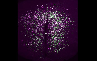
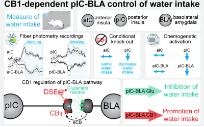
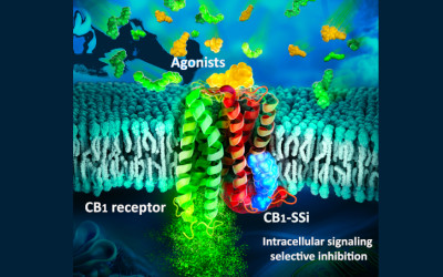
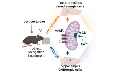

01/2012 | Nat Neurosci
Moving bliss: a new anandamide transporter.
Marsicano G, Chaouloff F
doi: 10.1038/nn.3011


2012 | Sci Rep
Developmental regulation of CB1-mediated spike-time dependent depression at immature mossy fiber-CA3 synapses.
Caiati MD, Sivakumaran S, Lanore F, Mulle C, Richard E, Verrier D, Marsicano G, Miles R, Cherubini E
doi: 10.1038/srep00285

Abstract:
Early in postnatal life, mossy fibres (MF), the axons of granule cells in the dentate gyrus, release GABA which is depolarizing and excitatory. Synaptic currents undergo spike-time dependent long-term depression (STD-LTD) regardless of the temporal order of stimulation (pre versus post and viceversa). Here we show that at P3 but not at P21, STD-LTD, induced by negative pairing, is mediated by endocannabinoids mobilized from the postsynaptic cell during spiking-induced membrane depolarization. By diffusing backward, endocannabinoids activate cannabinoid type-1 (CB1) receptors probably expressed on MF. Thus, STD-LTD was prevented by CB1 receptor antagonists and was absent in CB1-KO mice. Consistent with these data, in situ hybridization experiments revealed detectable level of CB1 mRNA in the granule cell layer at P3 but not at P21. These results indicate that CB1 receptors are transiently expressed on immature MF terminals where they counteract the enhanced neuronal excitability induced by the excitatory action of GABA.


2012 | PLoS ONE
Endocannabinoids measurement in human saliva as potential biomarker of obesity.
Matias I, Cherifi-Gatta B, Tabarin A, Clark S, Leste-Lasserre T, Marsicano G, Piazza PV, Cota D
doi: 10.1371/journal.pone.0042399

Abstract:
BACKGROUND: The discovery of the endocannabinoid system and of its role in the regulation of energy balance has significantly advanced our understanding of the physiopathological mechanisms leading to obesity and type 2 diabetes. New knowledge on the role of this system in humans has been acquired by measuring blood endocannabinoids. Here we explored endocannabinoids and related N-acylethanolamines in saliva and verified their changes in relation to body weight status and in response to a meal or to body weight loss. METHODOLOGY/PRINCIPAL FINDINGS: Fasting plasma and salivary endocannabinoids and N-acylethanolamines were measured through liquid mass spectrometry in 12 normal weight and 12 obese, insulin-resistant subjects. Salivary endocannabinoids and N-acylethanolamines were evaluated in the same cohort before and after the consumption of a meal. Changes in salivary endocannabinoids and N-acylethanolamines after body weight loss were investigated in a second group of 12 obese subjects following a 12-weeks lifestyle intervention program. The levels of mRNAs coding for enzymes regulating the metabolism of endocannabinoids, N-acylethanolamines and of cannabinoid type 1 (CB(1)) receptor, alongside endocannabinoids and N-acylethanolamines content, were assessed in human salivary glands. The endocannabinoids 2-arachidonoylglycerol (2-AG), N-arachidonoylethanolamide (anandamide, AEA), and the N-acylethanolamines (oleoylethanolamide, OEA and palmitoylethanolamide, PEA) were quantifiable in saliva and their levels were significantly higher in obese than in normal weight subjects. Fasting salivary AEA and OEA directly correlated with BMI, waist circumference and fasting insulin. Salivary endocannabinoids and N-acylethanolamines did not change in response to a meal. CB(1) receptors, ligands and enzymes were expressed in the salivary glands. Finally, a body weight loss of 5.3% obtained after a 12-weeks lifestyle program significantly decreased salivary AEA levels. CONCLUSIONS/SIGNIFICANCE: Endocannabinoids and N-acylethanolamines are quantifiable in saliva and their levels correlate with obesity but not with feeding status. Body weight loss significantly decreases salivary AEA, which might represent a useful biomarker in obesity.


16/11/2011 | J Neurosci
State-dependent, bidirectional modulation of neural network activity by endocannabinoids.
Piet R, Garenne A, Farrugia F, Le Masson G, Marsicano G, Chavis P, Manzoni OJ
doi: 10.1523/JNEUROSCI.4297-11.2011

Abstract:
The endocannabinoid (eCB) system and the cannabinoid CB1 receptor (CB1R) play key roles in the modulation of brain functions. Although actions of eCBs and CB1Rs are well described at the synaptic level, little is known of their modulation of neural activity at the network level. Using microelectrode arrays, we have examined the role of CB1R activation in the modulation of the electrical activity of rat and mice cortical neural networks in vitro. We find that exogenous activation of CB1Rs expressed on glutamatergic neurons decreases the spontaneous activity of cortical neural networks. Moreover, we observe that the net effect of the CB1R antagonist AM251 inversely correlates with the initial level of activity in the network: blocking CB1Rs increases network activity when basal network activity is low, whereas it depresses spontaneous activity when its initial level is high. Our results reveal a complex role of CB1Rs in shaping spontaneous network activity, and suggest that the outcome of endogenous neuromodulation on network function might be state dependent.


04/2011 | Physiology (Bethesda)
Endocannabinoids and motor behavior: CB1 receptors also control running activity.
Chaouloff F, Dubreucq S, Bellocchio L, Marsicano G
doi: 10.1152/physiol.00050.2010


Abstract:
Activation of both presynaptic metabotropic cannabinoid type 1 receptors (CB(1)s) and ionotropic kainate receptors (KARs) can efficiently modulate GABA release at many synapses of the CNS. The inhibitory effect of kainic acid (KA) has been ascribed to metabotropic actions, and KAR-induced release of secondary neuromodulatory agents may partly mediate these actions. Here, we investigated the involvement of the endocannabinoid system in the modulation of GABAergic synaptic transmission by pharmacological activation of KARs with KA in CA1 pyramidal neurons of the mouse hippocampus. We show that the depression of GABAergic synaptic transmission induced by KA (3 mum) is strongly inhibited by the simultaneous blockade of CB(1) and GABA(B) receptors with SR141716A (5 mum) and CGP55845 (5 mum), respectively. KA induces a calcium-dependent mobilization of the endocannabinoid anandamide (AEA) by activation of GluK2-containing KARs in postsynaptic pyramidal neurons. Consistently, the effect of KA is prolonged by the inhibitor of AEA degradation URB597 (1 mum) in a CB(1)-dependent manner, but it is not altered by blockade of degradation or synthesis of the other main endocannabinoid 2-arachidonoylglycerol (2AG). Hence, our work reveals that the pharmacological activation of KARs leads to the stimulation of secondary metabotropic signaling systems. In addition, these data further underline the profound mechanistic differences between exogenous and endogenous activation of KARs in the hippocampus.


02/2011 | J Neurophysiol
Presynaptic CB1 receptors regulate synaptic plasticity at cerebellar parallel fiber synapses.
Carey MR, Myoga MH, McDaniels KR, Marsicano G, Lutz B, Mackie K, Regehr WG
doi: 10.1152/jn.00980.2010

Abstract:
Endocannabinoids are potent regulators of synaptic strength. They are generally thought to modify neurotransmitter release through retrograde activation of presynaptic type 1 cannabinoid receptors (CB1Rs). In the cerebellar cortex, CB1Rs regulate several forms of synaptic plasticity at synapses onto Purkinje cells, including presynaptically expressed short-term plasticity and, somewhat paradoxically, a postsynaptic form of long-term depression (LTD). Here we have generated mice in which CB1Rs were selectively eliminated from cerebellar granule cells, whose axons form parallel fibers. We find that in these mice, endocannabinoid-dependent short-term plasticity is eliminated at parallel fiber, but not inhibitory interneuron, synapses onto Purkinje cells. Further, parallel fiber LTD is not observed in these mice, indicating that presynaptic CB1Rs regulate long-term plasticity at this synapse.


Abstract:
There is overwhelming evidence for multiple effects of stress on excitatory transmission and synaptic plasticity in the hippocampus. These interactions between stress and hippocampal glutamatergic neurons play a role in the cognitive and emotional consequences of aversive stimuli. Stress impacts on excitatory synapses are mediated by a complex set of neurohormones and neurotransmitters, among which corticosteroid hormones secreted from the adrenal cortex play a crucial role. Most effects of corticosteroid hormones are mediated by their binding to cytosolic mineralocorticoid (MR) and glucocorticoid receptors (GR), which after translocation to the nucleus, regulate the transcription of target genes. Recent electrophysiological and live imaging experiments have however provided experimental data which reinforce the hypothesis that beside these delayed effects, corticosteroid hormones may also act rapidly through membrane receptors. The first goal of this review is to detail the tonic and intrinsic effects of corticosteroid hormones on hippocampal excitatory transmission, glutamate receptor trafficking and expression, and synaptic plasticity, paying attention to their temporality (rapid and transient effects followed by slow and persistent genomic effects). Its second goal is to dissect the extent to which acute/repeated stress influences hippocampal excitatory synapses and whether these are accounted for by corticosteroid hormones.


01/2011 | Brain
Loss of striatal type 1 cannabinoid receptors is a key pathogenic factor in Huntington's disease.
Blazquez C, Chiarlone A, Sagredo O, Aguado T, Pazos MR, Resel E, Palazuelos J, Julien B, Salazar M, Borner C, Benito C, Carrasco C, Diez-Zaera M, Paoletti P, Diaz-Hernandez M, Ruiz C, Sendtner M, Lucas JJ, de Yebenes JG, Marsicano G, Monory K, Lutz B, Romero J, Alberch J, Gines S, Kraus J, Fernandez-Ruiz J, Galve-Roperh I, Guzman M
doi: 10.1093/brain/awq278

Abstract:
Endocannabinoids act as neuromodulatory and neuroprotective cues by engaging type 1 cannabinoid receptors. These receptors are highly abundant in the basal ganglia and play a pivotal role in the control of motor behaviour. An early downregulation of type 1 cannabinoid receptors has been documented in the basal ganglia of patients with Huntington's disease and animal models. However, the pathophysiological impact of this loss of receptors in Huntington's disease is as yet unknown. Here, we generated a double-mutant mouse model that expresses human mutant huntingtin exon 1 in a type 1 cannabinoid receptor-null background, and found that receptor deletion aggravates the symptoms, neuropathology and molecular pathology of the disease. Moreover, pharmacological administration of the cannabinoid Delta(9)-tetrahydrocannabinol to mice expressing human mutant huntingtin exon 1 exerted a therapeutic effect and ameliorated those parameters. Experiments conducted in striatal cells show that the mutant huntingtin-dependent downregulation of the receptors involves the control of the type 1 cannabinoid receptor gene promoter by repressor element 1 silencing transcription factor and sensitizes cells to excitotoxic damage. We also provide in vitro and in vivo evidence that supports type 1 cannabinoid receptor control of striatal brain-derived neurotrophic factor expression and the decrease in brain-derived neurotrophic factor levels concomitant with type 1 cannabinoid receptor loss, which may contribute significantly to striatal damage in Huntington's disease. Altogether, these results support the notion that downregulation of type 1 cannabinoid receptors is a key pathogenic event in Huntington's disease, and suggest that activation of these receptors in patients with Huntington's disease may attenuate disease progression.


2011 | PLoS ONE
GABAergic and cortical and subcortical glutamatergic axon terminals contain CB1 cannabinoid receptors in the ventromedial nucleus of the hypothalamus.
Reguero L, Puente N, Elezgarai I, Mendizabal-Zubiaga J, Canduela MJ, Buceta I, Ramos A, Suarez J, Rodriguez de Fonseca F, Marsicano G, Grandes P
doi: 10.1371/journal.pone.0026167

Abstract:
BACKGROUND: Type-1 cannabinoid receptors (CB(1)R) are enriched in the hypothalamus, particularly in the ventromedial hypothalamic nucleus (VMH) that participates in homeostatic and behavioral functions including food intake. Although CB(1)R activation modulates excitatory and inhibitory synaptic transmission in the brain, CB(1)R contribution to the molecular architecture of the excitatory and inhibitory synaptic terminals in the VMH is not known. Therefore, the aim of this study was to investigate the precise subcellular distribution of CB(1)R in the VMH to better understand the modulation exerted by the endocannabinoid system on the complex brain circuitries converging into this nucleus. METHODOLOGY/PRINCIPAL FINDINGS: Light and electron microscopy techniques were used to analyze CB(1)R distribution in the VMH of CB(1)R-WT, CB(1)R-KO and conditional mutant mice bearing a selective deletion of CB(1)R in cortical glutamatergic (Glu-CB(1)R-KO) or GABAergic neurons (GABA-CB(1)R-KO). At light microscopy, CB(1)R immunolabeling was observed in the VMH of CB(1)R-WT and Glu-CB(1)R-KO animals, being remarkably reduced in GABA-CB(1)R-KO mice. In the electron microscope, CB(1)R appeared in membranes of both glutamatergic and GABAergic terminals/preterminals. There was no significant difference in the percentage of CB(1)R immunopositive profiles and CB(1)R density in terminals making asymmetric or symmetric synapses in CB(1)R-WT mice. Furthermore, the proportion of CB(1)R immunopositive terminals/preterminals in CB(1)R-WT and Glu-CB(1)R-KO mice was reduced in GABA-CB(1)R-KO mutants. CB(1)R density was similar in all animal conditions. Finally, the percentage of CB(1)R labeled boutons making asymmetric synapses slightly decreased in Glu-CB(1)R-KO mutants relative to CB(1)R-WT mice, indicating that CB(1)R was distributed in cortical and subcortical excitatory synaptic terminals. CONCLUSIONS/SIGNIFICANCE: Our anatomical results support the idea that the VMH is a relevant hub candidate in the endocannabinoid-mediated modulation of the excitatory and inhibitory neurotransmission of cortical and subcortical pathways regulating essential hypothalamic functions for the individual's survival such as the feeding behavior.


Abstract:
Reactive Oxygen Species (ROS) constitute important intracellular signaling molecules. Mitochondria are admitted sources of ROS, especially of superoxide anions through the electron transport chain. Here the mitochondria-targeted ratiometric pericam (RPmt) was used as a superoxide biosensor, by appropriate choice of the excitation wavelength. RPmt was transfected in vivo into mouse muscles. Confocal imaging of isolated muscle fibers reveals spontaneous flashes of RPmt fluorescence. Flashes correspond to increases in superoxide production, as shown by simultaneous recordings of the fluorescence from MitoSox, a mitochondrial superoxide probe. Flashes occur in all subcellular populations of mitochondria. Spatial analysis of the flashes pattern over time revealed that arrays of mitochondria work as well-defined superoxide-production-units. Increase of superoxide production at the muscle fiber level involves recruitment of supplemental units with no increase in per-unit production. Altogether, these results demonstrate that superoxide flashes in muscle fibers correspond to physiological signals linked to mitochondrial metabolism. They also suggest that superoxide, or one of its derivatives, modulates its own production at the mitochondrial level.


09/2010 | j gen physiol
Paradoxical buffering of calcium by calsequestrin demonstrated for the calcium store of skeletal muscle.
Royer L, Sztretye M, Manno C, Pouvreau S, Zhou J, Knollmann BC, Protasi F, Allen PD, Rios E
doi: 10.1085/jgp.201010454

Abstract:
Contractile activation in striated muscles requires a Ca(2+) reservoir of large capacity inside the sarcoplasmic reticulum (SR), presumably the protein calsequestrin. The buffering power of calsequestrin in vitro has a paradoxical dependence on [Ca(2+)] that should be valuable for function. Here, we demonstrate that this dependence is present in living cells. Ca(2+) signals elicited by membrane depolarization under voltage clamp were compared in single skeletal fibers of wild-type (WT) and double (d) Casq-null mice, which lack both calsequestrin isoforms. In nulls, Ca(2+) release started normally, but the store depleted much more rapidly than in the WT. This deficit was reflected in the evolution of SR evacuability, E, which is directly proportional to SR Ca(2+) permeability and inversely to its Ca(2+) buffering power, B. In WT mice E starts low and increases progressively as the SR is depleted. In dCasq-nulls, E started high and decreased upon Ca(2+) depletion. An elevated E in nulls is consistent with the decrease in B expected upon deletion of calsequestrin. The different value and time course of E in cells without calsequestrin indicate that the normal evolution of E reflects loss of B upon SR Ca(2+) depletion. Decrement of B upon SR depletion was supported further. When SR calcium was reduced by exposure to low extracellular [Ca(2+)], release kinetics in the WT became similar to that in the dCasq-null. E became much higher, similar to that of null cells. These results indicate that calsequestrin not only stores Ca(2+), but also varies its affinity in ways that progressively increase the ability of the store to deliver Ca(2+) as it becomes depleted, a novel feedback mechanism of potentially valuable functional implications. The study revealed a surprisingly modest loss of Ca(2+) storage capacity in null cells, which may reflect concurrent changes, rather than detract from the physiological importance of calsequestrin.


01/08/2010 | J Physiol
In vivo expression of G-protein beta1gamma2 dimer in adult mouse skeletal muscle alters L-type calcium current and excitation-contraction coupling.
Weiss N, Legrand C, Pouvreau S, Bichraoui H, Allard B, Zamponi GW, De Waard M, Jacquemond V
doi: 10.1113/jphysiol.2010.191593

Abstract:
A number of G-protein-coupled receptors are expressed in skeletal muscle but their roles in muscle physiology and downstream effector systems remain poorly investigated. Here we explored the functional importance of the G-protein betagamma (Gbetagamma) signalling pathway on voltage-controlled Ca(2+) homeostasis in single isolated adult skeletal muscle fibres. A GFP-tagged Gbeta(1)gamma(2) dimer was expressed in vivo in mice muscle fibres. The GFP fluorescence pattern was consistent with a Gbeta(1)gamma(2) dimer localization in the transverse-tubule membrane. Membrane current and indo-1 fluorescence measurements performed under voltage-clamp conditions reveal a drastic reduction of both L-type Ca(2+) current density and of peak amplitude of the voltage-activated Ca(2+) transient in Gbeta(1)gamma(2)-expressing fibres. These effects were not observed upon expression of Gbeta(2)gamma(2), Gbeta(3)gamma(2) or Gbeta(4)gamma(2). Our data suggest that the G-protein beta(1)gamma(2) dimer may play an important regulatory role in skeletal muscle excitation-contraction coupling.


Abstract:
Chronic voluntary wheel-running activity has been reported to hypersensitise central CB1 receptors in mice. On the other hand, pharmacological findings suggest that the CB1 receptor could be involved in wheel-running behaviour and in running-induced neurogenesis in the hippocampus. We analysed wheel-running behaviour for 6 weeks and measured its consequences on hippocampal neurogenesis in CB1 knockout (CB1(-/-)) animals, compared to wild-type (CB1(+/+)) littermates. Because wheel running has been shown to affect locomotor reactivity in novel environments, memory for aversive events and depression-like behaviours, we also assessed these behaviours in control and running CB1(+/+) and CB1(-/-) mice. When compared with running CB1(+/+) mice, the distance covered weekly by CB1(-/-) mice was decreased by 30-40%, an observation accounted for by decreased time spent and maximal velocity on the wheels. Analyses of running distances with respect to the light/dark cycle revealed that mutant covered less distance throughout both the inactive and the active phases of that cycle. Locomotion in an activity cage, exploration in an open field, and immobility time in the forced swim test proved insensitive to chronic wheel running in either genotype. Wheel running, per se, did not influence the expression and extinction of cued fear memory but counteracted in a time-dependent manner the deficiency of extinction measured in CB1(-/-) mice. Hippocampal neurogenesis, assessed by doublecortin labelling of immature neurons in the dentate gyrus, was lowered by 40% in control CB1(-/-) mice, compared to control CB1(+/+) mice. Although CB1(-/-) mice ran less than their wild-type littermates, the 6-week running protocol increased neurogenesis to similar extents (37-39%) in both genotypes. This study suggests that mouse CB1 receptors control wheel running but not its neurogenic consequences in the hippocampus.


07/04/2010 | Cell Metab
CB(1) signaling in forebrain and sympathetic neurons is a key determinant of endocannabinoid actions on energy balance
Quarta C, Bellocchio L, Mancini G, Mazza R, Cervino C, Braulke LJ, Fekete C, Latorre R, Nanni C, Bucci M, Clemens LE, Heldmaier G, Watanabe M, Leste-Lasserre T, Maitre M, Tedesco L, Fanelli F, Reuss S, Klaus S, Srivastava RK, Monory K, Valerio A, Grandis A, De Giorgio R, Pasquali R, Nisoli E, Cota D, Lutz B, Marsicano G, Pagotto U
doi: 10.1016/j.cmet.2010.02.015


07/04/2010 | Cell Metab
CB(1) signaling in forebrain and sympathetic neurons is a key determinant of endocannabinoid actions on energy balance.
Quarta C, Bellocchio L, Mancini G, Mazza R, Cervino C, Braulke LJ, Fekete C, Latorre R, Nanni C, Bucci M, Clemens LE, Heldmaier G, Watanabe M, Leste-Lassere T, Maitre M, Tedesco L, Fanelli F, Reuss S, Klaus S, Srivastava RK, Monory K, Valerio A, Grandis A, De Giorgio R, Pasquali R, Nisoli E, Cota D, Lutz B, Marsicano G, Pagotto U
doi: 10.1016/j.cmet.2010.02.015

Abstract:
The endocannabinoid system (ECS) plays a critical role in obesity development. The pharmacological blockade of cannabinoid receptor type 1 (CB(1)) has been shown to reduce body weight and to alleviate obesity-related metabolic disorders. An unsolved question is at which anatomical level CB(1) modulates energy balance and the mechanisms involved in its action. Here, we demonstrate that CB(1) receptors expressed in forebrain and sympathetic neurons play a key role in the pathophysiological development of diet-induced obesity. Conditional mutant mice lacking CB(1) expression in neurons known to control energy balance, but not in nonneuronal peripheral organs, displayed a lean phenotype and resistance to diet-induced obesity. This phenotype results from an increase in lipid oxidation and thermogenesis as a consequence of an enhanced sympathetic tone and a decrease in energy absorption. In conclusion, CB(1) signaling in the forebrain and sympathetic neurons is a key determinant of the ECS control of energy balance.


03/2010 | Nat Neurosci
Bimodal control of stimulated food intake by the endocannabinoid system
Bellocchio L*, Lafenetre P*, Cannich A, Cota D, Puente N, Grandes P, Chaouloff F, Piazza PV*, Marsicano G*
doi: 10.1038/nn.2494


03/2010 | Pflugers Arch
Altered myoplasmic Ca(2+) handling in rat fast-twitch skeletal muscle fibres during disuse atrophy.
Weiss N, Andrianjafiniony T, Dupre-Aucouturier S, Pouvreau S, Desplanches D, Jacquemond V
doi: 10.1007/s00424-009-0764-x

Abstract:
Calcium-dependent signalling pathways are believed to play an important role in skeletal muscle atrophy, but whether intracellular Ca(2+) homeostasis is affected in that situation remains obscure. We show here that there is a 20% atrophy of the fast-type flexor digitorum brevis (FDB) muscle in rats hind limb unloaded (HU) for 2 weeks, with no change in fibre type distribution. In voltage-clamp experiments, the amplitude of the slow Ca(2+) current was found similar in fibres from control and HU animals. In fibres loaded with the Ca(2+) dye indo-1, the value for the rate of [Ca(2+)] decay after the end of 5-100-ms-long voltage-clamp depolarisations from -80 to +10 mV was found to be 30-50% lower in fibres from HU animals. This effect was consistent with a reduced contribution of both saturable and non-saturable components of myoplasmic Ca(2+) removal. However, there was no change in the relative amount of parvalbumin, and type 1 sarco-endoplasmic reticulum Ca(2+)-ATPase was increased by a factor of three in the atrophied muscles. Confocal imaging of mitochondrial membrane potential showed that atrophied FDB fibres had significantly depolarized mitochondria as compared to control fibres. Depolarization of mitochondria in control fibres with carbonyl cyanide-p-trifluoromethoxyphenylhydrazone induced a slowing of the decay of [Ca(2+)] transients accompanied by an increase in resting [Ca(2+)] and a reduction of the peak amplitude of the transients. Overall results provide the first functional evidence for severely altered intracellular Ca(2+) removal capabilities in atrophied fast-type muscle fibres and highlight the possible contribution of reduced mitochondrial polarisation.


02/2010 | Nat Neurosci
Synaptic activation of kainate receptors gates presynaptic CB(1) signaling at GABAergic synapses
Lourenco J, Cannich A, Carta M, Coussen F, Mulle C*, Marsicano G*
doi: 10.1038/nn.2481


25/01/2010 | PLoS ONE
Localization and function of the cannabinoid CB1 receptor in the anterolateral bed nucleus of the stria terminalis
Puente N, Elezgarai I, Lafourcade M, Reguero L, Marsicano G, Georges F, Manzoni O*, Grandes P*
doi: 10.1371/journal.pone.0008869


Abstract:
The balance between novelty seeking and safety assessment is a key feature of adaptive behavior, and alterations in this equilibrium can lead to neuropsychiatric disorders. Excessive novelty seeking is a main form of pathological impulsivity, which is among the symptoms that define attention deficit hyperactivity disorder (ADHD). There is growing evidence that the endocannabinoid system (ECS) plays an important role in the control of this balance, but little is known about the underlying neuronal mechanisms. In this study, we aimed at dissecting the neurocircuits under the control of the ECS in novelty-induced behavioral inhibition. To reach this goal, we combined pharmacological, genetic and behavioral tools. Mice were repeatedly exposed to novel palatable food or a novel object and their responses to these stimuli were analyzed over several days. The results confirmed that systemic blockade of cannabinoid type-1 (CB(1)) receptors strongly decreases palatable food intake, but its impact onto the response to novelty is less pronounced. Using conditional mutant mice lacking the CB(1) receptor either in cortical glutamatergic or in GABAergic neurons, we found that the ECS exerts opposite functions on the balance between novelty seeking and behavioral inhibition. Whereas CB(1) receptors expressed in cortical glutamatergic neurons favors novelty seeking, CB(1)-dependent control of inhibitory GABAergic neurons promotes behavioral inhibition. These data show a tightly regulated influence of the ECS on impulsive behaviors and suggest the involvement of endocannabinoid signaling in the pathophysiological modulation of ADHD and related disorders.


12/2009 | Nat Neurosci
Self-modulation of neocortical pyramidal neurons by endocannabinoids.
Marinelli S, Pacioni S, Cannich A, Marsicano G, Bacci A
doi: 10.1038/nn.2430

Abstract:
Control of pyramidal neuron excitability is vital for the functioning of the neocortex. Somatodendritic slow self-inhibition (SSI) allows inhibitory neurons to regulate their own activity, but the existence of similar mechanisms in excitatory cells has not been shown. We found that in rodents endocannabinoids mediated SSI and long-term modulation of inhibitory connections in layer 2/3 pyramidal neurons with a distinct dendritic morphology, suggesting that a glutamatergic network in cortical circuits is self-regulated.


Abstract:
To investigate the impact of averseness, controllability and familiarity of a test situation on the involvement of the endocannabinoid system in the regulation of exploratory behaviour, we tested conventional and conditional cannabinoid receptor type 1 (CB1)-deficient mice in behavioural paradigms with different emotional load, which depended on the strength of illumination and the ability of the animals to avoid the light stimulus. Complete CB1 null-mutant mice (Total-CB1-KO) showed an anxiogenic-like phenotype under circumstances where they were able to avoid the bright light such as the elevated plus-maze and the light/dark avoidance task. Conditional mutant mice lacking CB1 expression specifically in cortical glutamatergic neurons (Glu-CB1-KO), in contrast, failed to show a similar phenotype under the same experimental conditions. However, both mutant lines showed increased avoidance of open arm exploration during a second exposure to the elevated plus-maze. If tested in situations where the fear eliciting light could not be avoided, Total-CB1-KO mice showed increased thigmotaxis in an open field, decreased social investigation and decreased novel object exploration under aversive light conditions, but not under non-aversive low light. This time, Glu-CB1-KO also showed decreased exploratory behaviour towards objects and conspecific juveniles and increased thigmotaxis in the open field. Taking into consideration that the behavioural performance of wild-type mice was only marginally affected by changes in light intensities, these data indicate that the endocannabinoid system renders exploratory behaviour largely independent of the test averseness. This process differentially involves endocannabinoid-controlled glutamatergic transmission, depending on the controllability of the test situation.


09/2009 | Nat Neurosci
Cannabinoid modulation of hippocampal long-term memory is mediated by mTOR signaling.
Puighermanal E, Marsicano G, Busquets-Garcia A, Lutz B, Maldonado R, Ozaita A
doi: 10.1038/nn.2369

Abstract:
Cognitive impairment is one of the most important negative consequences associated with cannabis consumption. We found that CB1 cannabinoid receptor (CB1R) activation transiently modulated the mammalian target of rapamycin (mTOR)/p70S6K pathway and the protein synthesis machinery in the mouse hippocampus, which correlated with the amnesic properties of delta9-tetrahydrocannabinol (THC). In addition, non-amnesic doses of either the mTOR blocker rapamycin or the protein synthesis inhibitor anisomycin abrogated the amnesic-like effects of THC, pointing to a mechanism involving new protein synthesis. Moreover, using pharmacological and genetic tools, we found that THC long-term memory deficits were mediated by CB1Rs expressed on GABAergic interneurons through a glutamatergic mechanism, as both the amnesic-like effects and p70S6K phosphorylation were reduced in GABA-CB1R knockout mice and by NMDA blockade.


07/08/2009 | Science
Spinal endocannabinoids and CB1 receptors mediate C-fiber-induced heterosynaptic pain sensitization.
Pernia-Andrade AJ, Kato A, Witschi R, Nyilas R, Katona I, Freund TF, Watanabe M, Filitz J, Koppert W, Schuttler J, Ji G, Neugebauer V, Marsicano G, Lutz B, Vanegas H, Zeilhofer HU
doi: 10.1126/science.1171870

Abstract:
Diminished synaptic inhibition in the spinal dorsal horn is a major contributor to chronic pain. Pathways that reduce synaptic inhibition in inflammatory and neuropathic pain states have been identified, but central hyperalgesia and diminished dorsal horn synaptic inhibition also occur in the absence of inflammation or neuropathy, solely triggered by intense nociceptive (C-fiber) input to the spinal dorsal horn. We found that endocannabinoids, produced upon strong nociceptive stimulation, activated type 1 cannabinoid (CB1) receptors on inhibitory dorsal horn neurons to reduce the synaptic release of gamma-aminobutyric acid and glycine and thus rendered nociceptive neurons excitable by nonpainful stimuli. Our results suggest that spinal endocannabinoids and CB1 receptors on inhibitory dorsal horn interneurons act as mediators of heterosynaptic pain sensitization and play an unexpected role in dorsal horn pain-controlling circuits.


06/2009 | Eur J Neurosci
WIN55,212-2, a cannabinoid receptor agonist, protects against nigrostriatal cell loss in the 1-methyl-4-phenyl-1,2,3,6-tetrahydropyridine mouse model of Parkinson's disease.
Price DA, Martinez AA, Seillier A, Koek W, Acosta Y, Fernandez E, Strong R, Lutz B, Marsicano G, Roberts JL, Giuffrida A
doi: 10.1111/j.1460-9568.2009.06764.x

Abstract:
Parkinson's disease (PD) is characterized by the progressive loss of nigrostriatal dopamine neurons leading to motor disturbances and cognitive impairment. Current pharmacotherapies relieve PD symptoms temporarily but fail to prevent or slow down the disease progression. In this study, we investigated the molecular mechanisms by which the non-selective cannabinoid receptor agonist WIN55,212-2 (WIN) protects mouse nigrostriatal neurons from 1-methyl-4-phenyl-1,2,3,6-tetrahydropyridine (MPTP)-induced neurotoxicity and neuroinflammation. Stereological analyses showed that chronic treatment with WIN (4 mg/kg, intraperitoneal), initiated 24 h after MPTP administration, protected against MPTP-induced loss of tyrosine hydroxylase-positive neurons in the substantia nigra pars compacta independently of CB1 cannabinoid receptor activation. The neuroprotective effect of WIN was accompanied by increased dopamine and 3,4-dihydroxyphenylacetic acid levels in the substantia nigra pars compacta and dorsal striatum of MPTP-treated mice. At 3 days post-MPTP, we found significant microglial activation and up-regulation of CB2 cannabinoid receptors in the ventral midbrain. Treatment with WIN or the CB2 receptor agonist JWH015 (4 mg/kg, intraperitoneal) reduced MPTP-induced microglial activation, whereas genetic ablation of CB2 receptors exacerbated MPTP systemic toxicity. Furthermore, chronic WIN reversed MPTP-associated motor deficits, as revealed by the analysis of forepaw step width and percentage of faults using the inverted grid test. In conclusion, our data indicate that agonism at CB2 cannabinoid receptors protects against MPTP-induced nigrostriatal degeneration by inhibiting microglial activation/infiltration and suggest that CB2 receptors represent a new therapeutic target to slow the degenerative process occurring in PD.


20/03/2009 | acs chem biol
Indo-1 derivatives for local calcium sensing.
Bannwarth M, Correa IR, Sztretye M, Pouvreau S, Fellay C, Aebischer A, Royer L, Rois E, Johnsson K
doi: 10.1021/cb800258g

Abstract:
The role of calcium in signal transduction relies on the precise spatial and temporal control of its concentration. The existing means to detect fluctuations in Ca2+ concentrations with adequate temporal and spatial resolution are limited. We introduce here a method to measure Ca2+ concentrations in defined locations in living cells that is based on linking the Ca2+-sensitive dye Indo-1 to SNAP-tag fusion proteins. Fluorescence spectroscopy of SNAP-Indo-1 conjugates in vitro showed that the conjugates retained the Ca2+-sensing ability of Indo-1. In a proof-of-principle experiment, local Ca2+ sensing was demonstrated in single cells dissociated from muscle of adult mice expressing a nucleus-localized SNAP-tag fusion. Ca2+ concentrations inside nuclei of resting cells were measured by shifted excitation and emission ratioing of confocal microscopic images of fluorescence. After permeabilizing the plasma membrane, changes in the bathing solution induced corresponding changes in nuclear [Ca2+] that were readily detected and used for a preliminary calibration of the technique. This work thus demonstrates the synthesis and application of SNAP-tag-based Ca2+ indicators that combine the spatial specificity of genetically encoded calcium indicators with the advantageous spectroscopic properties of synthetic indicators.


Abstract:
Recent evidence showed that the endocannabinoid system plays an important role in the behavioral adaptation of stress and fear responses. In this study, we chose a behavioral paradigm that includes criteria of both fear and stress responses to assess whether the involvement of endocannabinoids in these two processes rely on common mechanisms. To this end, we delivered a footshock and measured the fear response to a subsequently presented novel tone stimulus. First, we exposed different groups of cannabinoid receptor type 1 (CB(1))-deficient mice (CB(1) (-/-)) and their wild-type littermates (CB(1) (+/+)) to footshocks of different intensities. Only application of an intense footshock resulted in a sustained fear response to the tone in CB(1) (-/-). Using the intense protocol, we next investigated whether endocannabinoids mediate their effects via an interplay with corticotropin-releasing hormone (CRH) signaling. Pharmacological blockade of CB(1) receptors by rimonabant in mice deficient for the CRH receptor type 1 (CRHR1(-/-)) or type 2 (CRHR2(-/-)), and in respective wild-type littermates, resulted in a sustained fear response in all genotypes. This suggests that CRH is not involved in the fear-alleviating effects of CB(1). As CRHR1(-/-) are known to be severely impaired in stress-induced corticosterone secretion, our observation also implicates that corticosterone is dispensable for CB(1)-mediated acute fear adaptation. Instead, conditional mutants with a specific deletion of CB(1) in principal neurons of the forebrain (CaMK-CB(1) (-/-)), or in cortical glutamatergic neurons (Glu-CB(1) (-/-)), showed a similar phenotype as CB(1) (-/-), thus indicating that endocannabinoid-controlled glutamatergic transmission plays an essential role in acute fear adaptation.


2009 | Science
Spinal endocannabinoids mediate activity-dependent central sensitization in pain pathways
Pernia-Andrade AJ, Witschi R, Nyials R, Katona I, Freund TF, Watanabe M, Filitz J, Koppert W, Schüttler J, Ji G, Neugebauer V, Marsicano G, Lutz B, Vanegas H, Zeilhofer HU


2009 | Eur J Neurosci
WO'55,212,2, a cannabinoid receptor agonist, protects against nigrostriatal cell loss in the 1-methyl-4-phenyl-1,2,3,4-tetrahydropyridine mouse model of Parkinson's disease.
Price DA, Martinez AA, Seillier A, Koek W, Acosta Y, Fernandez H, Strong R, Lutz B, Marsicano G, Roberts JL, Giuffrida A


2009 | Curr Top Behav Neurosci
Roles of the endocannabinoid system in learning and memory.
Marsicano G, Lafenetre P
doi: 10.1007/978-3-540-88955-7_8

Abstract:
The endocannabinoid system (ECS) plays a central role in the regulation of learning and memory processes. The fine-tuned regulation of neural transmission by the system is likely to be the mechanism underlying this important function. In this chapter, we review the data in the literature showing the direct involvement of the physiological activation of cannabinoid receptors in the modulation of different forms of learning and memory. When possible, we also address the likely mechanisms of this involvement. Finally, given the apparent special role of the ECS in the extinction of fear, we propose a reasonable model to assess how neuronal networks could be influenced by the endocannabinoids in these processes. Overall, the data reviewed indicate that, despite the enormous progress of recent years, much is still to be done to fully elucidate the mechanisms of the ECS influence on learning and memory processes.


Abstract:
The elementary Ca(2+)-release events underlying voltage-activated myoplasmic Ca(2+) transients in mammalian muscle remain elusive. Here, we looked for such events in confocal line-scan (x,t) images of fluo-3 fluorescence taken from isolated adult mouse skeletal muscle fibers held under voltage-clamp conditions. In response to step depolarizations, spatially segregated fluorescence signals could be detected that were riding on a global increase in fluorescence. These discrete signals were separated using digital filtering in the spatial domain; mean values for their spatial half-width and amplitude were 1.99 +/- 0.09 microm and 0.16 +/- 0.005 DeltaF/F(0) (n = 151), respectively. Under control conditions, the duration of the events was limited by the pulse duration. In contrast, in the presence of maurocalcine, a scorpion toxin suspected to disrupt the process of repolarization-induced ryanodine receptor (RyR) closure, events uninterrupted by the end of the pulse were readily detected. Overall results establish these voltage-activated low-amplitude local Ca(2+) signals as inherent components of the physiological Ca(2+)-release process of mammalian muscle and suggest that they result from the opening of either one RyR or a coherently operating group of RyRs, under the control of the plasma membrane polarization.


Abstract:
Intracellular calcium signals regulate multiple cellular functions. They depend on release of Ca(2+) from cellular stores into the cytosol, a process that in many types of cells appears to be tightly controlled by changes in [Ca(2+)] within the store. In contrast with cardiac muscle, where depletion of Ca(2+) in the sarcoplasmic reticulum is a crucial determinant of termination of Ca(2+) release, in skeletal muscle there is no agreement regarding the sign, or even the existence of an effect of SR Ca(2+) level on Ca(2+) release. To address this issue we measured Ca(2+) transients in mouse flexor digitorum brevis (FDB) skeletal muscle fibres under voltage clamp, using confocal microscopy and the Ca(2+) monitor rhod-2. The evolution of Ca(2+) release flux was quantified during long-lasting depolarizations that reduced severely the Ca(2+) content of the SR. As in all previous determinations in mammals and non-mammals, release flux consisted of an early peak, relaxing to a lower level from which it continued to decay more slowly. Decay of flux in this second stage, which has been attributed largely to depletion of SR Ca(2+), was studied in detail. A simple depletion mechanism without change in release permeability predicts an exponential decay with time. In contrast, flux decreased non-exponentially, to a finite, measurable level that could be maintained for the longest pulses applied (1.8 s). An algorithm on the flux record allowed us to define a quantitative index, the normalized flux rate of change (NFRC), which was shown to be proportional to the ratio of release permeability P and inversely proportional to Ca(2+) buffering power B of the SR, thus quantifying the 'evacuability' or ability of the SR to empty its content. When P and B were constant, flux then decayed exponentially, and NFRC was equal to the exponential rate constant. Instead, in most cases NFRC increased during the pulse, from a minimum reached immediately after the early peak in flux, to a time between 200 and 250 ms, when the index was no longer defined. NFRC increased by 111% on average (in 27 images from 18 cells), reaching 300% in some cases. The increase may reflect an increase in P, a decrease in B, or both. On experimental and theoretical grounds, both changes are to be expected upon SR depletion. A variable evacuability helps maintain a constant Ca(2+) output under conditions of diminishing store Ca(2+) load.


09/2008 | J Clin Invest
Hepatic CB1 receptor is required for development of diet-induced steatosis, dyslipidemia, and insulin and leptin resistance in mice
Osei-Hyiaman D, Liu J, Zhou L, Godlewski G, Harvey-White J, Jeong W I, Batkai S, Marsicano G, Lutz B, Buettner C, Kunos G
doi: 10.1172/JCI34827

Abstract:
Diet-induced obesity is associated with fatty liver, insulin resistance, leptin resistance, and changes in plasma lipid profile. Endocannabinoids have been implicated in the development of these associated phenotypes, because mice deficient for the cannabinoid receptor CB1 (CB1-/-) do not display these changes in association with diet-induced obesity. The target tissues that mediate these effects, however, remain unknown. We therefore investigated the relative role of hepatic versus extrahepatic CB1 receptors in the metabolic consequences of a high-fat diet, using liver-specific CB1 knockout (LCB1-/-) mice. LCB1(-/-) mice fed a high-fat diet developed a similar degree of obesity as that of wild-type mice, but, similar to CB1(-/-) mice, had less steatosis, hyperglycemia, dyslipidemia, and insulin and leptin resistance than did wild-type mice fed a high-fat diet. CB1 agonist-induced increase in de novo hepatic lipogenesis and decrease in the activity of carnitine palmitoyltransferase-1 and total energy expenditure were absent in both CB1(-/-) and LCB1(-/-) mice. We conclude that endocannabinoid activation of hepatic CB1 receptors contributes to the diet-induced steatosis and associated hormonal and metabolic changes, but not to the increase in adiposity, observed with high-fat diet feeding. Theses studies suggest that peripheral CB1 receptors could be selectively targeted for the treatment of fatty liver, impaired glucose homeostasis, and dyslipidemia in order to minimize the neuropsychiatric side effects of nonselective CB1 blockade during treatment of obesity-associated conditions.


Abstract:
The complete genetic loss or pharmacological blockade of cannabinoid receptor type 1 (CB1) in mice results in both altered behavioral performance and increased stress hormone secretion in response to stressful encounters such as forced swim test (FST) exposure. CB1 is expressed on nerve terminals belonging to different neurotransmitter systems, including the glutamatergic and GABAergic system, where it is able to suppress excitatory and inhibitory neurotransmission, respectively. In the current study, we used the conditional mutagenesis approach in mice to investigate the neurotransmitter systems involved in these behavioral and neuroendocrine phenotypes in regard to CB1 signaling. Mice lacking CB1 in cortical glutamatergic neurons (Glu-CB1(-/-)) showed decreased passive stress coping (decreased immobility) in the FST, whereas mice lacking CB1 in principal forebrain neurons (CaMK-CB1(-/-)) and GABAergic neurons (GABA-CB1(-/-)), respectively, behaved as littermate controls. However, we found increased FST-induced corticosterone secretion only in CaMK-CB1(-/-) mice, whereas Glu-CB1(-/-) and GABA-CB1(-/-) mice exhibited normal corticosterone release as compared to controls. Thus, behavioral and neuroendocrine acute stress coping in response to the FST is mainly influenced by CB1 signaling on different glutamatergic neuronal subpopulations, but not by CB1 on GABAergic neurons.


Abstract:
Corticosterone facilitates hippocampal glutamate transmission, but the cellular pathways by which AMPA receptor (AMPAR) signaling is adjusted remain elusive. Single quantum-dot imaging in live rat hippocampal neurons revealed that corticosterone triggers, via distinct corticosteroid receptors, time-dependent increases in GluR2-AMPAR surface mobility and synaptic surface GluR2 content. Furthermore, corticosterone potentiates the increase of synaptic surface GluR2 contents by a chemical long-term potentiation stimulus, revealing the influence that corticosterone has on AMPAR trafficking.