Publications


IF du Neurocentre
IF1234567891011121314151617181920253035404550
Nombre1217571601251078156373217171812312271211811116719
%11386543221111211111111111


2221 publications


25/11/2024 | Mol Metab
TGR5 receptors in SF1-expressing neurons of the ventromedial hypothalamus regulate glucose homeostasis.
Zizzari P, Castellanos-Jankiewicz A, Yagoub S, Simon V, Clark S, Maître M, Dupuy N, Leste-Lasserre T, Gonzales D, Schoonjans K, Fénelon VS, Cota D
doi: 10.1016/j.molmet.2024.102071

Abstract:
OBJECTIVE: Steroidogenic factor-1 (SF1) neurons of the ventromedial hypothalamus play key roles in the regulation of food intake, body weight and glucose metabolism. The bile acid receptor Takeda G protein-coupled receptor 5 (TGR5) is expressed in the hypothalamus, where it determines some of the actions of bile acids on food intake and body weight through still poorly defined neuronal mechanisms. Here, we examined the role of TGR5 in SF1 neurons in the regulation of energy balance and glucose metabolism. METHODS: We used a genetic approach combined with metabolic phenotyping and molecular analyses to establish the effect of TGR5 deletion in SF1 neurons on meal pattern, body weight, body composition, energy expenditure and use of energy substrates as well as on possible changes in glucose handling and insulin sensitivity. RESULTS: Our findings reveal that TGR5 in SF1 neurons does not play a major role in the regulation of food intake or body weight under standard chow, but it is involved in the adaptive feeding response to the acute exposure to cold or to a hypercaloric, high-fat diet, without changes in energy expenditure. Notably, TGR5 in SF1 neurons hinder glucose metabolism, since deletion of the receptor improves whole-body glucose uptake through heightened insulin signaling in the hypothalamus and in the brown adipose tissue. CONCLUSIONS: TGR5 in SF1 neurons favours satiety by differently modifying the meal pattern in response to specific metabolic cues. These studies also reveal a novel key function for TGR5 in SF1 neurons in the regulation of whole-body insulin sensitivity, providing new insight into the role played by neuronal TGR5 in the regulation of metabolism.


10/10/2024 | j neuroinflammation
Astrocytic DLL4-NOTCH1 signaling pathway promotes neuroinflammation via the IL-6-STAT3 axis.
Mora P, Laisne M, Bourguignon C, Rouault P, Jaspard-Vinassa B, Maitre M, Gadeau AP, Renault MA, Horng S, Couffinhal T, Chapouly C
doi: 10.1186/s12974-024-03246-w

Abstract:
Under neuroinflammatory conditions, astrocytes acquire a reactive phenotype that drives acute inflammatory injury as well as chronic neurodegeneration. We hypothesized that astrocytic Delta-like 4 (DLL4) may interact with its receptor NOTCH1 on neighboring astrocytes to regulate astrocyte reactivity via downstream juxtacrine signaling pathways. Here we investigated the role of astrocytic DLL4 on neurovascular unit homeostasis under neuroinflammatory conditions. We probed for downstream effectors of the DLL4-NOTCH1 axis and targeted these for therapy in two models of CNS inflammatory disease. We first demonstrated that astrocytic DLL4 is upregulated during neuroinflammation, both in mice and humans, driving astrocyte reactivity and subsequent blood-brain barrier permeability and inflammatory infiltration. We then showed that the DLL4-mediated NOTCH1 signaling in astrocytes directly drives IL-6 levels, induces STAT3 phosphorylation promoting upregulation of astrocyte reactivity markers, pro-permeability factor secretion and consequent blood-brain barrier destabilization. Finally we revealed that blocking DLL4 with antibodies improves experimental autoimmune encephalomyelitis symptoms in mice, identifying a potential novel therapeutic strategy for CNS autoimmune demyelinating disease. As a general conclusion, this study demonstrates that DLL4-NOTCH1 signaling is not only a key pathway in vascular development and angiogenesis, but also in the control of astrocyte reactivity during neuroinflammation.


09/10/2024 | org biomol chem
Synthesis and biological evaluation of natural Lachnophyllum methyl ester, Lachnophyllum lactone and their synthetic analogs.
Adande K, Simalou O, Ardanuy J, Eloh K, Mehalla C, Constant P, Fabing I, Genisson Y, Ballereau S

Abstract:
(2Z)-Lachnophyllum methyl ester and (4Z)-Lachnophyllum lactone were recently identified as major components in essential oils and extracts of Conyza bonariensis from Togo. Extended biological evaluation of these acetylenic compounds was however hampered by the reduced amounts isolated. A synthetic route was designed providing access to larger quantities of these two natural products as well as to original non-natural analogs with the prospect of exploring for the first time the structure-activity relationships in this series. Using LC/MS analysis, synthetic samples allowed confirming the presence of the two previously isolated natural products in plant extracts obtained by the accelerated solvent extraction technique. The nematocidal activity of the synthesized compounds confirmed the potency of the natural products, which remain the most active among all analogs tested. The synthesized compounds were also assessed against Leishmania infantum axenic amastigotes and the Mycobacterium tuberculosis H(37)Rv pathogenic strain. (2Z)-Lachnophyllum methyl ester, (4Z)-Lachnophyllum lactone and lactone analogs exhibited the strongest antileishmanial potency. As expected, a longer alkyl chain was necessary to observe significant antimycobacterial activity. The lactone analog bearing a C10 lipophilic appendage displayed the highest antimycobacterial potency. The notable activities of lactones, naturally occurring or analogs, either nematicidal, antileishmanial or antimycobacterial, were compared to their cytotoxicity for mammalian cells and revealed moderate selectivity index values. In this regard, the innocuous (2Z)-Lachnophyllum methyl ester and its analogs open up more promising perspectives for the discovery of bioactive agents to protect both agricultural crops and human health.


10/2024 | BioRxiv
Dopamine transmission in the anterior insula shapes the neural coding of anxiety
Couderc Y, Dhanireddy T, Vardiero G, Garg A, Ricci D, D'almeida M, Nicolas C, Habchi T, Wu KY, Gjorgjieva J, Li Y, Valjent E, Beyeler A
doi: https://doi.org/10.1101/2024.10.25.620186

Abstract:
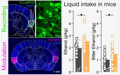
The insular cortex (or insula), and particularly its anterior region, plays a crucial role in the control of emotional valence and anxiety (Etkin & Wager, 2007; Méndez-Ruette et al., 2019; Nicolas et al., 2023). While dopamine neurotransmission is known to modulate anxiety levels in humans (Hjorth et al., 2021) and animal models (de la Mora et al., 2010; Bananej et al., 2012; Zarrindast & Khakpai, 2015; DeGroot et al., 2020; Godino et al., 2023), its regulatory effects on the anterior insula remained unexplored. Here, using a multifaceted approach, we uncovered how dopamine shapes anterior insula function in anxiety and valence processing. First, we revealed a high density of neurons expressing type-1 dopamine receptors (D1) in the insula, particularly important in the anterior insula, and seven times greater than the density of neurons expressing type-2 dopamine receptors (D2). Few neurons co-expressed Drd1 and Drd2 mRNAs in the anterior and posterior insula, and the density of Drd1+ neurons in the anterior insula was twice higher among inhibitory neurons than excitatory neurons. Second, we found that pharmacological activation of D1 in the anterior insula is anxiogenic, suggesting a direct link between insular dopamine signaling and anxiety-related behaviors. Using fiber-photometry recordings, we identified that the amplitude of dopamine release onto D1+ neurons in the anterior insula while mice were in anxiogenic spaces or receiving mild foot shocks was both positively correlated with mice level of trait anxiety. Population dynamics and deep-learning analyses of anterior insula single-unit recordings uncovered distinct coding patterns of anxiety-provoking and safe environments, as well as tastants of positive and negative valence. Remarkably, systemic D1 activation, which heightens anxiety- related behaviors, dampens this coding dichotomy by increasing coding variability for protected spaces while increasing the coding reliability for anxiogenic spaces. Interestingly, the coding reliability of anxiogenic areas was positively correlated with mice level of trait anxiety, and we observed a trend towards a positive correlation between the coding reliability of a negative tastants, and mice level of anxiety. Altogether, our findings provide a new model of neural population coding of anxiety and emotional valence and unravel D1-dependent coding mechanisms in the mouse anterior insula.



25/09/2024 | Int J Biochem Cell Biol
Classical psychedelics' action on brain monoaminergic systems.
Butler JJ, Ricci D, Aman C, Beyeler A, De Deurwaerdere P

Abstract:
The study of the mechanism of action of classical psychedelics has gained significant interest due to their clinical potential in the treatment of several psychiatric conditions, including major depressive and anxiety disorders. These drugs bind 5-hydroxytryptamine receptors (5-HTR) including 5-HT(1A)R, 5-HT(2A)R, 5-HT(2B)R, and/or 5-HT(2)(C)R, as well as other targets. 5-HTRs regulate the activity of ascending monoaminergic neurons, a mechanism primarily involved in the action of classical antidepressant drugs, antipsychotics, and drugs of abuse. Sparse neurochemical data have been produced on the control of monoaminergic neuron activity in response to classical psychedelics. Here we review the available data in order to determine whether classical psychedelics have specific neurochemical effects on serotonergic, dopaminergic, and noradrenergic neurons. The data show that these drugs have disparate effects on each monoaminergic system, demonstrating a complex response with state-dependent and region-specific effects. For instance, several psychedelics inhibit the firing of serotonergic neurons, although this is not necessarily associated with a decrease in serotonin release in all regions. Noradrenergic neuron spontaneous activity also appears to be inhibited by psychedelics, also not necessarily associated with a decrease in noradrenaline release in all regions. Psychedelics influence on dopaminergic systems is also complex as the above-mentioned 5-HTRs may have opposing effects on dopaminergic neuron activity, in a state-dependent manner. There is an apparent lack of clear neuronal signature induced by psychedelics on monoaminergic neuron activity despite specific recurrent mechanisms. This review provides a current summary of the action of psychedelics on monoamine neuromodulators serotonin, dopamine and noradrenaline, compiling reoccurring and contradictory findings demonstrating that a monoamine signature of psychedelics, if applicable, would be state- and region-dependant.


14/09/2024 | Prog Neuropsychopharmacol Biol Psychiatry
Sequential physical and cognitive training disrupts cocaine-context associations via multi-level stimulation of adult hippocampal neurogenesis.
Ávila-Gámiz F, Pérez-Cano AM, Pérez-Berlanga JM, Zambrana-Infantes EN, Mañas-Padilla MC, Gil-Rodríguez S, Tronel S, Santín LJ, Ladrón de Guevara-Miranda D
doi: 10.1016/j.pnpbp.2024.111148

Abstract:
Cocaine-related contextual cues are a recurrent source of craving and relapse. Extinction of cue-driven cocaine seeking remains a clinical challenge, and the search for adjuvants is ongoing. In this regard, combining physical and cognitive training is emerging as a promising strategy that has shown synergistic benefits on brain structure and function, including enhancement of adult hippocampal neurogenesis (AHN), which has been recently linked to reduced maintenance of maladaptive drug seeking. Here, we examined whether this behavioral approach disrupts cocaine-context associations via improved AHN. To this aim, C57BL/6J mice (N = 37) developed a cocaine-induced conditioned place preference (CPP) and underwent interventions consisting of exercise and/or spatial working memory training. Bromodeoxyuridine (BrdU) was administered during early running sessions to tag a subset of new dentate granule cells (DGCs) reaching a critical window of survival during spatial learning. Once these DGCs became functionally mature (∼ 6 weeks-old), mice received extinction training before testing CPP extinction and reinstatement. We found that single and combined treatments accelerated CPP extinction and prevented reinstatement induced by a low cocaine priming (2 mg/kg). Remarkably, the dual-intervention mice showed a significant decrease of CPP after extinction relative to untreated animals. Moreover, combining the two strategies led to increased number and functional integration of BrdU(+) DGCs, which in turn maximized the effect of spatial training (but not exercise) to reduce CPP persistence. Together, our findings suggests that sequencing physical and cognitive training may redound to decreased maintenance of cocaine-context associations, with multi-level stimulation of AHN as a potential underlying mechanism.


12/09/2024 | adv healthc mater
Dye-Based Fluorescent Organic Nanoparticles, New Promising Tools for Optogenetics.
Lesas J, Bienvenu TCM, Kurek E, Verlhac JB, Grivet Z, Têtu M, Girard D, Lanore F, Blanchard-Desce M, Herry C, Daniel J, Dejean C
doi: 10.1002/adhm.202402132

Abstract:
Dye-based fluorescent organic nanoparticles are a specific class of nanoparticles obtained by nanoprecipitation in water of pure dyes only. While the photophysical and colloidal properties of the nanoparticles strongly depend on the nature of the aggregated dyes, their excellent brightness in the visible and in the near infrared make these nanoparticles a unique and versatile platform for in vivo application. This article examines the promising utilization of these nanoparticles for in vivo optogenetics applications. Their photophysical properties as well as their biocompatibility and their capacity to activate Chrimson opsin in vivo through the fluorescence reabsorption process are demonstrated. Additionally, an illustrative example of employing these nanoparticles in fear reduction in mice through closed-loop stimulation is presented. Through an optogenetic methodology, the nanoparticles demonstrate an ability to selectively manipulate neurons implicated in the fear response and diminish the latter. Dye-based fluorescent organic nanoparticles represent a promising and innovative strategy for optogenetic applications, holding substantial potential in the domain of translational neuroscience. This work paves the way for novel therapeutic modalities for neurological and neuropsychiatric disorders.


Abstract:
Microglia, as the resident macrophages of the brain, are essential for maintaining brain homeostasis. They shape neuronal circuits during development, survey their environment for debris or dead cells, as well as respond to infection and injury in the brain, among many other functions. However, their important role in neurodevelopment and synaptic plasticity and pathophysiology has not been fully defined, highlighting the need for further investigation. To gain a more comprehensive understanding of the role of microglia in these processes, we need to isolate microglia and characterize them genetically, metabolically, and functionally. However, the isolation of microglia from adult mice, especially from small brain structures, is challenging as they represent a small percentage of the total brain cells, and the yield of isolated microglia is often too low. Here, the magnetic isolation of microglia using CD11b(+) microbeads allows us to sort microglial cells from the hypothalamus of a freshly perfused adult mouse brain. The current method allows us to achieve relatively high purity and yield in a short period while maintaining cell viability.


04/09/2024 | Nat Commun
Cannabidiol ameliorates mitochondrial disease via PPARgamma activation in preclinical models.
Puighermanal E, Luna-Sanchez M, Gella A, van der Walt G, Urpi A, Royo M, Tena-Morraja P, Appiah I, de Donato MH, Menardy F, Bianchi P, Esteve-Codina A, Rodriguez-Pascau L, Vergara C, Gomez-Pallares M, Marsicano G, Bellocchio L, Martinell M, Sanz E, Jurado S, Soriano FX, Pizcueta P, Quintana A

Abstract:
Mutations in mitochondrial energy-producing genes lead to a heterogeneous group of untreatable disorders known as primary mitochondrial diseases (MD). Leigh syndrome (LS) is the most common pediatric MD and is characterized by progressive neuromuscular affectation and premature death. Here, we show that daily cannabidiol (CBD) administration significantly extends lifespan and ameliorates pathology in two LS mouse models, and improves cellular function in fibroblasts from LS patients. CBD delays motor decline and neurodegenerative signs, improves social deficits and breathing abnormalities, decreases thermally induced seizures, and improves neuropathology in affected brain regions. Mechanistically, we identify peroxisome proliferator-activated receptor gamma (PPARgamma) as a key nuclear receptor mediating CBD's beneficial effects, while also providing proof of dysregulated PPARgamma expression and activity as a common feature in both mouse neurons and fibroblasts from LS patients. Taken together, our results provide the first evidence for CBD as a potential treatment for LS.


21/08/2024 | J Neurosci
GAP43 located on corticostriatal terminals restrains novelty-induced hyperactivity in mice.
Maroto IB, Costas-Insua C, Montero-Fernandez C, Hermoso-Lopez A, Lebouc M, Bajo-Graneras R, Alvaro-Blazquez A, Blazquez C, Cannich A, Marsicano G, Martin R, Baufreton J, Rodriguez-Crespo I, Bellocchio L, Guzman M

Abstract:
Growth-associated protein of 43 kDa (GAP43) is a key cytoskeleton-associated component of the presynaptic terminal that facilitates neuroplasticity. Downregulation of GAP43 expression has been associated to various psychiatric conditions in humans and evokes hippocampus-dependent memory impairments in mice. Despite the extensive studies conducted on hippocampal GAP43 in past decades, however, very little is known about its roles in modulating the excitatory vs. inhibitory balance in other brain regions. We recently generated conditional knockout mice in which the Gap43 gene was selectively inactivated in either telencephalic glutamatergic neurons (Gap43(fl/fl) ;Nex1(Cre) mice, hereafter Glu-GAP43(-/-) mice) or forebrain GABAergic neurons (Gap43(fl/fl) ;Dlx5/6(Cre) mice, hereafter GABA-GAP43(-/-) mice). Here, we show that Glu-GAP43(-/-) but not GABA-GAP43(-/-) mice of either sex show a striking hyperactive phenotype when exposed to a novel environment. This behavioral alteration of Glu-GAP43(-/-) mice was linked to a selective activation of dorsal-striatum neurons, as well as to an enhanced corticostriatal glutamatergic transmission and an abrogation of corticostriatal endocannabinoid-mediated long-term depression. In line with these observations, GAP43 was abundantly expressed in corticostriatal glutamatergic terminals of wild-type mice. The novelty-induced hyperactive phenotype of Glu-GAP43(-/-) mice was abrogated by chemogenetically inhibiting corticostriatal afferences with a G(i)-coupled 'designer receptor exclusively activated by designer drugs' (DREADD), thus further supporting that novelty-induced activity is controlled by GAP43 at corticostriatal excitatory projections. Taken together, these findings show an unprecedented regulatory role of GAP43 in the corticostriatal circuitry and provide a new mouse model with a delimited neuronal-circuit alteration for studying novelty-induced hyperactivity, a phenotypic shortfall that occurs in diverse psychiatric diseases.Significance statement Psychiatric alterations such as attention deficit/hyperactivity disorder, schizophrenia and bipolar disorder pose a significant health and socioeconomic burden to our society. Animal models that recapitulate precise phenotypical traits of those diseases are therefore warranted for developing new therapeutic interventions. Here, we found that mice lacking the protein GAP43 selectively in telencephalic glutamatergic neurons show a robust novelty-induced hyperactive phenotype, a behavioral deficit often associated to psychiatric diseases. These mice exhibit profound alterations in corticostriatal excitatory plasticity and a selective overactivation of dorsal-striatum neurons in response to a novel environment. Our findings thus unveil an important role of GAP43 in corticostriatal function and provide a new animal model with a delimited neuronal-circuit alteration for studying novelty-induced hyperactivity in psychiatric disorders.


18/08/2024 | Nat Commun
Olfactory bulb astrocytes link social transmission of stress to cognitive adaptation in male mice.
Gomez-Sotres P, Skupio U, Dalla Tor T, Julio-Kalajzic F, Cannich A, Gisquet D, Bonilla-Del Rio I, Drago F, Puente N, Grandes P, Bellocchio L, Busquets-Garcia A, Bains JS, Marsicano G
doi: 10.1038/s41467-024-51416-4

Abstract:
Emotions and behavior can be affected by social chemosignals from conspecifics. For instance, olfactory signals from stressed individuals induce stress-like physiological and synaptic changes in naive partners. Direct stress also alters cognition, but the impact of socially transmitted stress on memory processes is currently unknown. Here we show that exposure to chemosignals produced by stressed individuals is sufficient to impair memory retrieval in unstressed male mice. This requires astrocyte control of information in the olfactory bulb mediated by mitochondria-associated CB1 receptors (mtCB1). Targeted genetic manipulations, in vivo Ca(2+) imaging and behavioral analyses reveal that mtCB1-dependent control of mitochondrial Ca(2+) dynamics is necessary to process olfactory information from stressed partners and to define their cognitive consequences. Thus, olfactory bulb astrocytes provide a link between social odors and their behavioral meaning.


13/08/2024 | Nat Commun
A neuronal circuit driven by GLP-1 in the olfactory bulb regulates insulin secretion.
Montaner M, Denom J, Simon V, Jiang W, Holt MK, Brierley DI, Rouch C, Foppen E, Kassis N, Jarriault D, Khan D, Eygret L, Mifsud F, Hodson DJ, Broichhagen J, Van Oudenhove L, Fioramonti X, Gault V, Cota D, Reimann F, Gribble FM, Migrenne-Li S, Trapp S, Gurden H, Magnan C

Abstract:
Glucagon-like peptide 1 (GLP-1) stimulates insulin secretion and holds significant pharmacological potential. Nevertheless, the regulation of energy homeostasis by centrally-produced GLP-1 remains partially understood. Preproglucagon cells, known to release GLP-1, are found in the olfactory bulb (OB). We show that activating GLP-1 receptors (GLP-1R) in the OB stimulates insulin secretion in response to oral glucose in lean and diet-induced obese male mice. This is associated with reduced noradrenaline content in the pancreas and blocked by an α(2)-adrenergic receptor agonist, implicating functional involvement of the sympathetic nervous system (SNS). Inhibiting GABA(A) receptors in the paraventricular nucleus of the hypothalamus (PVN), the control centre of the SNS, abolishes the enhancing effect on insulin secretion induced by OB GLP-1R. Therefore, OB GLP-1-dependent regulation of insulin secretion relies on a relay within the PVN. This study provides evidence that OB GLP-1 signalling engages a top-down neural mechanism to control insulin secretion via the SNS.


09/08/2024 | Nat Commun
A lactate-dependent shift of glycolysis mediates synaptic and cognitive processes in male mice.
Fernandez-Moncada I, Lavanco G, Fundazuri UB, Bollmohr N, Mountadem S, Dalla Tor T, Hachaguer P, Julio-Kalajzic F, Gisquet D, Serrat R, Bellocchio L, Cannich A, Fortunato-Marsol B, Nasu Y, Campbell RE, Drago F, Cannizzaro C, Ferreira G, Bouzier-Sore AK, Pellerin L, Bolanos JP, Bonvento G, Barros LF, Oliet SHR, Panatier A, Marsicano G
doi: 10.1038/s41467-024-51008-2

Abstract:
Astrocytes control brain activity via both metabolic processes and gliotransmission, but the physiological links between these functions are scantly known. Here we show that endogenous activation of astrocyte type-1 cannabinoid (CB1) receptors determines a shift of glycolysis towards the lactate-dependent production of D-serine, thereby gating synaptic and cognitive functions in male mice. Mutant mice lacking the CB1 receptor gene in astrocytes (GFAP-CB1-KO) are impaired in novel object recognition (NOR) memory. This phenotype is rescued by the gliotransmitter D-serine, by its precursor L-serine, and also by lactate and 3,5-DHBA, an agonist of the lactate receptor HCAR1. Such lactate-dependent effect is abolished when the astrocyte-specific phosphorylated-pathway (PP), which diverts glycolysis towards L-serine synthesis, is blocked. Consistently, lactate and 3,5-DHBA promoted the co-agonist binding site occupancy of CA1 post-synaptic NMDA receptors in hippocampal slices in a PP-dependent manner. Thus, a tight cross-talk between astrocytic energy metabolism and gliotransmission determines synaptic and cognitive processes.


07/08/2024 | Nat Commun
A plastic aggrecan barrier modulated by peripheral energy state gates metabolic signal access to arcuate neurons.
Kuczynski-Noyau L, Karmann S, Alberton P, Martinez-Corral I, Nampoothiri S, Sauvé F, Lhomme T, Quarta C, Apte SS, Bouret S, Aszodi A, Rasika S, Ciofi P, Dam J, Prévot V, Mattot V
doi: 10.1038/s41467-024-50798-9

Abstract:
The hypothalamic arcuate nucleus (ARH) contains neurons vital for maintaining energy homeostasis that sense and respond to changes in blood-borne metabolic hormones. Despite its juxtaposition to the median eminence (ME), a circumventricular organ lacking a blood-brain barrier and thus exposed to circulating molecules, only a few ventral ARH neurons perceive these extravasating metabolic signals due to a poorly understood ME/ARH diffusion barrier. Here, we show in male mice that aggrecan, a perineural-net proteoglycan deposited by orexigenic ARH neurons, creates a peculiar ventrodorsal diffusion gradient. Fasting enhances aggrecan deposition more dorsally, reinforcing the diffusion barrier, particularly around neurons adjacent to fenestrated capillary loops that enter the ARH. The disruption of aggrecan deposits results in unregulated diffusion of blood-borne molecules into the ARH and impairs food intake. Our findings reveal the molecular nature and plasticity of the ME/ARH diffusion barrier, and indicate its physiological role in hypothalamic metabolic hormone sensing.


Abstract:
Amyloid-peptide (Abeta) monomeric forms (ABM) occurring in presymptomatic Alzheimer's disease (AD) brain are thought to be devoid of neurotoxicity while the transition/aggregation of ABM into oligomers is determinant for Abeta-induced toxicity since Abeta is predominantly monomeric up to 3 microM and aggregates over this concentration. However, recent imaging and/or histopathological investigations revealed alterations of myelin in prodromal AD brain in absence of aggregated Abeta oligomers, suggesting that ABM may induce toxicity in myelin-producing cells in early AD-stages. To check this hypothesis, here we studied ABM effects on the viability of the Human oligodendrocyte cell line (HOG), a reliable oligodendrocyte model producing myelin proteins. Furthermore, to mimic closely interactions between oligodendrocytes and other glial cells regulating myelination, we investigated also ABM effects on mouse brain primary mixed-glial cell cultures. Various methods were combined to show that ABM concentrations (600 nM-1 microM), extremely lower than 3 microM, significantly decreased HOG cell and mouse brain primary mixed-glial cell survival. Interestingly, flow-cytometry studies using specific cell-type markers demonstrated that oligodendrocytes represent the most vulnerable glial cell population affected by ABM toxicity. Our work also shows that the neurosteroid 3alpha-O-allyl-allopregnanolone BR351 (250 and 500 nM) efficiently prevented ABM-induced HOG and brain primary glial cell toxicity. Bicuculline (50-100 nM), the GABA-A-receptor antagonist, was unable to block/reduce BR351 effect against ABM-induced HOG and primary glial cell toxicity, suggesting that BR351-evoked neuroprotection of these cells may not depend on GABA-A-receptor allosterically modulated by neurosteroids. Altogether, our results suggest that further exploration of BR351 therapeutic potential may offer interesting perspectives to develop effective neuroprotective strategies.


08/2024 | jbmr plus
Osteocyte gene expression analysis in mouse bone: optimization of a laser-assisted microdissection protocol.
Palmier M, Maitre M, Doat H, Leste-Lasserre T, Maurel DB, Boiziau C
doi: 10.1093/jbmrpl/ziae078

Abstract:
Among bone cells, osteocytes are the most abundant, but also the most challenging to study because they are located inside a dense mineralized matrix. Due to their involvement in bone homeostasis, diverse tools are needed to understand their roles in bone physiology and pathology. This work was aimed at establishing a laser-assisted microdissection protocol to isolate osteocytes and analyze their gene expressions. The goal was to overcome the limitations of the technique currently most used: RNA extraction from the whole bone. To perform laser microdissection and subsequent gene expression analysis, the five main steps of the protocol have been adapted for the bone tissue. After testing many parameters, we found that the best options were (1) take unfixed snap-frozen tissue, (2) cryosection with a supported tape system to improve the tissue morphology if necessary, (3) microdissect regions of interest, and (4) recover the bone pieces by catapulting, if feasible, or by gravity. Finally, RNA extraction (5) was the most efficient with a precipitation method and allowed quantifying the expression of well described osteocyte genes (Gja1/Cx43, Phex, Pdpn, Dmp1, Sost). This work describes two protocols optimized for femur and calvaria and gives an overview of the many optimization options that one could try when facing difficulties with laser microdissection.


30/07/2024 | J Am Coll Cardiol
Human DNA Extraction and Sequencing From Cardiomyocytes Collected by Catheter-Based Aspiration.
Maury P, Ader F, Lhuillier E, Martins F, Beneyto M, Gales C, Villard E, Duboscq-Bidot L, Gandjbakhch E, Guilbeau-Frugier C
doi: 10.1016/j.jacc.2024.05.037


21/06/2024 | npj microgravity
Physical exercise restores adult neurogenesis deficits induced by simulated microgravity.
Gros A, Furlan FM, Rouglan V, Favereaux A, Bontempi B, Morel JL
doi: 10.1038/s41526-024-00411-6

Abstract:
Cognitive impairments have been reported in astronauts during spaceflights and documented in ground-based models of simulated microgravity (SMG) in animals. However, the neuronal causes of these behavioral effects remain largely unknown. We explored whether adult neurogenesis, known to be a crucial plasticity mechanism supporting memory processes, is altered by SMG. Adult male Long-Evans rats were submitted to the hindlimb unloading model of SMG. We studied the proliferation, survival and maturation of newborn cells in the following neurogenic niches: the subventricular zone (SVZ)/olfactory bulb (OB) and the dentate gyrus (DG) of the hippocampus, at different delays following various periods of SMG. SMG exposure for 7 days, but not shorter periods of 6 or 24 h, resulted in a decrease of newborn cell proliferation restricted to the DG. SMG also induced a decrease in short-term (7 days), but not long-term (21 days), survival of newborn cells in the SVZ/OB and DG. Physical exercise, used as a countermeasure, was able to reverse the decrease in newborn cell survival observed in the SVZ and DG. In addition, depending on the duration of SMG periods, transcriptomic analysis revealed modifications in gene expression involved in neurogenesis. These findings highlight the sensitivity of adult neurogenesis to gravitational environmental factors during a transient period, suggesting that there is a period of adaptation of physiological systems to this new environment.


Abstract:
The dynamic suppression of threat-related behavior as a function of environmental constraint is critical for survival in mammals, yet the neurobiological underpinnings remain largely unknown. In this issue of Neuron, Wang et al.(1) identified prefrontal dynorphin-expressing neurons as key elements for tracking threat-related behavioral states and regulating fear suppression.


Abstract:
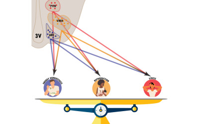
BACKGROUND: Recent advances in neuroscience tools for single-cell molecular profiling of brain neurons have revealed an enormous spectrum of neuronal subpopulations within the neuroendocrine hypothalamus, highlighting the remarkable molecular and cellular heterogeneity of this brain area. RATIONALE: Neuronal diversity in the hypothalamus reflects the high functional plasticity of this brain area, where multiple neuronal populations flexibly integrate a variety of physiological outputs, including energy balance, stress and fertility, through crosstalk mechanisms with peripheral hormones. Intrinsic functional heterogeneity is also observed within classically 'defined' subpopulations of neuroendocrine neurons, including subtypes with distinct neurochemical signatures, spatial organisation and responsiveness to hormonal cues. AIM: The aim of this review is to critically evaluate past and current research on the functional diversity of hypothalamic neuroendocrine neurons and their plasticity. It focuses on how this neuronal plasticity in this brain area relates to metabolic control, feeding regulation and interactions with stress and fertility-related neural circuits. CONCLUSION: Our analysis provides an original framework for improving our understanding of the hypothalamic regulation of hormone function and the development of neuroendocrine diseases.


Abstract:
Single administration of low-dose ketamine has both acute and sustained anti-depressant effects. Sustained effect is associated with restoration of glutamatergic synapses in medial prefrontal cortic (mFPC) neurons. Ketamine induced profound changes in a number of molecular pathways in a mouse model for chronic stress. Cell-cell communication analyses predicted that planar-cell-polarity (PCP) signaling was decreased after chronic administration of corticosterone but increased following ketamine administration in most of the excitatory neurons. Similar decrease of PCP signaling in excitatory neurons was predicted in dorsolateral prefrontal cortical (dl-PFC) neurons of patients with major depressive disorder (MDD). We showed that the basolateral amygdala (BLA)-projecting infralimbic prefrontal cortex (IL PFC) neurons regulate immobility time in the tail suspension test and food consumption. Conditionally knocking out Celsr2 and Celsr3 or Prickle2 in the BLA-projecting IL PFC neurons abolished ketamine-induced synapse restoration and behavioral remission. Therefore, PCP proteins in IL PFC-BLA neurons mediate synapse restoration induced by of low-dose ketamine.


03/06/2024 | J Pathol
Integrated spatial and multimodal single-cell transcriptomics reveal patient-dependent cell heterogeneity in splenic marginal zone lymphoma.
Cerapio JP, Gravelle P, Quillet-Mary A, Valle C, Martins F, Franchini DM, Syrykh C, Brousset P, Traverse-Glehen A, Ysebaert L, Fournie JJ, Laurent C
doi: 10.1002/path.6296

Abstract:
Biological hallmarks of splenic marginal zone lymphoma (SMZL) remain poorly described. Herein, we performed in-depth SMZL characterization through multimodal single-cell analyses of paired blood/spleen samples. The 3'-single-cell RNA-sequencing, Cellular Indexing of Transcriptomes and Epitopes by sequencing, and 5'-V(D)J single-cell RNA-sequencing datasets were integrated to characterize SMZL transcriptome profiles, including B-cell receptor and T-cell receptor repertoires. Hyperexpanded B-cell clones in the spleen were at a memory-like stage, whereas recirculating tumor B-cells in blood encompassed multiple differentiation stages, indicating an unexpected desynchronization of the B-cell maturation program in SMZL cells. Spatial transcriptomics showed the enrichment of T-effector and T-follicular helper (T(FH)) signatures in the nodular subtype of SMZL. This latter also exhibited gene-based cell-cell interactions suggestive of dynamic crosstalk between T(FH) and cancer cells in transcriptomics, further substantiated by using imaging mass cytometry. Our findings provide a comprehensive high-resolution description of SMZL biological hallmarks and characterize, for the first time in situ, inter- and intra-patient heterogeneity at both transcriptomic and protein levels. © 2024 The Authors. The Journal of Pathology published by John Wiley & Sons Ltd on behalf of The Pathological Society of Great Britain and Ireland.


Abstract:
Among the several animal models of alpha-synucleinopathies, the well-known viral vector-mediated delivery of wild-type or mutated (A53T) alpha-synuclein requires new tools to increase the lesion in mice and follow up in vivo expression. To this end, we developed a bioluminescent expression reporter of the human A53T-alpha-synuclein gene using the NanoLuc system into an AAV2/9, embedded or not in a fibroin solution to stabilise its expression in space and time. We first verified the expression of the fused protein in vitro on transfected cells by bioluminescence and Western blotting. Next, two groups of C57Bl6Jr mice were unilaterally injected with the AAV-NanoLuc-human-A53T-alpha-synuclein above the substantia nigra combined (or not) with fibroin. We first show that the in vivo cerebral bioluminescence signal was more intense in the presence of fibroin. Using immunohistochemistry, we find that the human-A53T-alpha-synuclein protein is more restricted to the ipsilateral side with an overall greater magnitude of the lesion when fibroin was added. However, we also detected a bioluminescence signal in peripheral organs in both conditions, confirmed by the presence of viral DNA corresponding to the injected AAV in the liver using qPCR.


24/04/2024 | Nat Commun
Single cell tracing of Pomc neurons reveals recruitment of 'Ghost' subtypes with atypical identity in a mouse model of obesity.
Leon S, Simon V, Lee TH, Steuernagel L, Clark S, Biglari N, Lesté-Lasserre T, Dupuy N, Cannich A, Bellocchio L, Zizzari P, Allard C, Gonzales D, Le Feuvre Y, Lhuillier E, Brochard A, Nicolas JC, Teillon J, Nikolski M, Marsicano G, Fioramonti X, Brüning JC, Cota D, Quarta C
doi: 10.1038/s41467-024-47877-2

Abstract:
The hypothalamus contains a remarkable diversity of neurons that orchestrate behavioural and metabolic outputs in a highly plastic manner. Neuronal diversity is key to enabling hypothalamic functions and, according to the neuroscience dogma, it is predetermined during embryonic life. Here, by combining lineage tracing of hypothalamic pro-opiomelanocortin (Pomc) neurons with single-cell profiling approaches in adult male mice, we uncovered subpopulations of 'Ghost' neurons endowed with atypical molecular and functional identity. Compared to 'classical' Pomc neurons, Ghost neurons exhibit negligible Pomc expression and are 'invisible' to available neuroanatomical approaches and promoter-based reporter mice for studying Pomc biology. Ghost neuron numbers augment in diet-induced obese mice, independent of neurogenesis or cell death, but weight loss can reverse this shift. Our work challenges the notion of fixed, developmentally programmed neuronal identities in the mature hypothalamus and highlight the ability of specialised neurons to reversibly adapt their functional identity to adult-onset obesogenic stimuli.


23/04/2024 | Cell Rep
Synaptic homeostasis transiently leverages Hebbian mechanisms for a multiphasic response to inactivity.
Sun SED, Levenstein D, Li B, Mandelberg N, Chenouard N, Suutari BS, Sanchez S, Tian G, Rinzel J, Buzsaki G, Tsien RW
doi: 10.1016/j.celrep.2024.113839

Abstract:
Homeostatic regulation of synapses is vital for nervous system function and key to understanding a range of neurological conditions. Synaptic homeostasis is proposed to operate over hours to counteract the destabilizing influence of long-term potentiation (LTP) and long-term depression (LTD). The prevailing view holds that synaptic scaling is a slow first-order process that regulates postsynaptic glutamate receptors and fundamentally differs from LTP or LTD. Surprisingly, we find that the dynamics of scaling induced by neuronal inactivity are not exponential or monotonic, and the mechanism requires calcineurin and CaMKII, molecules dominant in LTD and LTP. Our quantitative model of these enzymes reconstructs the unexpected dynamics of homeostatic scaling and reveals how synapses can efficiently safeguard future capacity for synaptic plasticity. This mechanism of synaptic adaptation supports a broader set of homeostatic changes, including action potential autoregulation, and invites further inquiry into how such a mechanism varies in health and disease.


15/04/2024 | Curr Biol
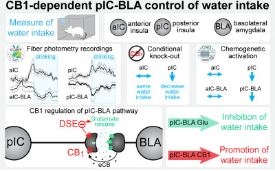
Cannabinoids regulate an insula circuit controlling water intake.
Zhao Z, Covelo A, Couderc Y, Mitra A, Varilh M, Wu Y, Jacky D, Fayad R, Cannich A, Bellocchio L, Marsicano G, Beyeler A
doi: 10.1016/j.cub.2024.03.053

Abstract:
The insular cortex, or insula, is a large brain region involved in the detection of thirst and the regulation of water intake. However, our understanding of the topographical, circuit, and molecular mechanisms for controlling water intake within the insula remains parcellated. We found that type-1 cannabinoid (CB(1)) receptors in the insular cortex cells participate in the regulation of water intake and deconstructed the circuit mechanisms of this control. Topographically, we revealed that the activity of excitatory neurons in both the anterior insula (aIC) and posterior insula (pIC) increases in response to water intake, yet only the specific removal of CB(1) receptors in the pIC decreases water intake. Interestingly, we found that CB(1) receptors are highly expressed in insula projections to the basolateral amygdala (BLA), while undetectable in the neighboring central part of the amygdala. Thus, we recorded the neurons of the aIC or pIC targeting the BLA (aIC-BLA and pIC-BLA) and found that they decreased their activity upon water drinking. Additionally, chemogenetic activation of pIC-BLA projection neurons decreased water intake. Finally, we uncovered CB(1)-dependent short-term synaptic plasticity (depolarization-induced suppression of excitation [DSE]) selectively in pIC-BLA, compared with aIC-BLA synapses. Altogether, our results support a model where CB(1) receptor signaling promotes water intake by inhibiting the pIC-BLA pathway, thereby contributing to the fine top-down control of thirst responses.


Abstract:
The development of 2D or 3D bioactive platforms for rapidly isolating pure populations of cells from adult stem cells holds promise for advancing the understanding of cellular mechanisms, drug testing, and tissue engineering. Over the years, methods have emerged to synthesize bioactive micro- and nanostructured 2D materials capable of directing stem cell fate. We introduce a novel method for randomly micro- or nanopatterning any protein/peptide onto both 2D and 3D scaffolds via spray technology. Our goal is to investigate the impact of arranging bioactive micropatterns (ordered vs disordered) on surfaces to guide human mesenchymal stem cell (hMSC) differentiation. The spray technology efficiently coats materials with controlled, cost-effective bioactive micropatterns in various sizes and shapes. BMP-2 mimetic peptides were covalently grafted, individually or in combination with RGD peptides, onto activated polyethylene terephthalate (PET) surfaces through a spraying process, incorporating nano/microscale parameters like size, shape, and composition. The study explores different peptide distributions on surfaces and various peptide combinations. Four surfaces were homogeneously functionalized with these peptides (M1 to M4 with various densities of peptides), and six surfaces with disordered micro- and nanopatterns of peptides (S0 to S5 with different sizes of peptide patterns) were synthesized. Fluorescence microscopy assessed peptide distribution, followed by hMSC culture for 2 weeks, and evaluated osteogenic differentiation via immunocytochemistry and RT-qPCR for osteoblast and osteocyte markers. Cells on uniformly peptide-functionalized surfaces exhibited cuboidal forms, while those on surfaces with disordered patterns tended toward columnar or cuboidal shapes. Surfaces S4 and S5 showed dendrite-like formations resembling an osteocyte morphology. S5 showed significant overexpression of osteoblast (OPN) and osteocyte markers (E11, DMP1, and SOST) compared to control surfaces and other micropatterned surfaces. Notably, despite sharing an equivalent quantity of peptides with a homogeneous functionalized surface, S5 displayed a distinct distribution of peptides, resulting in enhanced osteogenic differentiation of hMSCs.


04/2024 | Obesity (Silver Spring)
Antiobesity effects of intestinal gluconeogenesis are mediated by the brown adipose tissue sympathetic nervous system.
Vily-Petit J, Soty-Roca M, Silva M, Micoud M, Evrard F, Bron C, Raffin M, Beiroa D, Nogueiras R, Roussel D, Gautier-Stein A, Rajas F, Cota D, Mithieux G
doi: 10.1002/oby.23985

Abstract:
OBJECTIVE: Intestinal gluconeogenesis (IGN), via the initiation of a gut-brain nervous circuit, accounts for the metabolic benefits linked to dietary proteins or fermentable fiber in rodents and has been positively correlated with the rapid amelioration of body weight after gastric bypass surgery in humans with obesity. In particular, the activation of IGN moderates the development of hepatic steatosis accompanying obesity. In this study, we investigated the specific effects of IGN on adipose tissue metabolism, independent of its induction by nutritional manipulation. METHODS: We used two transgenic mouse models of suppression or overexpression of G6pc1, the catalytic subunit of glucose-6 phosphatase, which is the key enzyme of endogenous glucose production specifically in the intestine. RESULTS: Under a hypercaloric diet, mice overexpressing IGN showed lower adiposity and higher thermogenic capacities than wild-type mice, featuring marked browning of white adipose tissue (WAT) and prevention of the whitening of brown adipose tissue (BAT). Sympathetic denervation restricted to BAT caused the loss of the antiobesity effects associated with IGN. Conversely, IGN-deficient mice exhibited an increase in adiposity under a standard diet, which was associated with decreased expression of markers of thermogenesis in both BAT and WAT. CONCLUSIONS: IGN is sufficient to activate the sympathetic nervous system and prevent the expansion and the metabolic alterations of BAT and WAT metabolism under a high-calorie diet, thereby preventing the development of obesity. These data increase knowledge of the mechanisms of weight reduction in gastric bypass surgery and pave the way for new approaches to prevent or cure obesity.


04/2024 | Nat Rev Endocrinol
mTORC1 in energy expenditure: consequences for obesity.
Allard C, Miralpeix C, López-Gambero AJ, Cota D
doi: 10.1038/s41574-023-00934-0

Abstract:
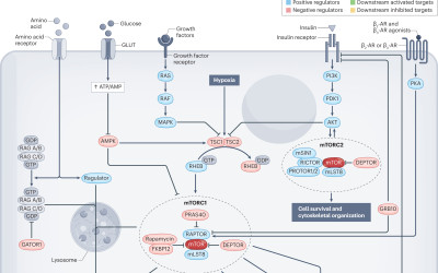
In eukaryotic cells, the mammalian target of rapamycin complex 1 (sometimes referred to as the mechanistic target of rapamycin complex 1; mTORC1) orchestrates cellular metabolism in response to environmental energy availability. As a result, at the organismal level, mTORC1 signalling regulates the intake, storage and use of energy by acting as a hub for the actions of nutrients and hormones, such as leptin and insulin, in different cell types. It is therefore unsurprising that deregulated mTORC1 signalling is associated with obesity. Strategies that increase energy expenditure offer therapeutic promise for the treatment of obesity. Here we review current evidence illustrating the critical role of mTORC1 signalling in the regulation of energy expenditure and adaptive thermogenesis through its various effects in neuronal circuits, adipose tissue and skeletal muscle. Understanding how mTORC1 signalling in one organ and cell type affects responses in other organs and cell types could be key to developing better, safer treatments targeting this pathway in obesity.


Abstract:
Spatial omics have been evolving rapidly in recent years, and due to the richness of the data they provide, they appear to offer opportunities for understanding biological phenomena and improving healthcare. Spatial omics enable the analysis of genomic, transcriptional, proteomic, and metabolic content at very high resolution. Beyond identifying molecular cell subpopulations, these approaches also provide information about the spatial location of the identified subpopulations within tissue, their proximity to each other, and their relationship with the extracellular matrix, blood vessels, and other tissue components. Spatial transcriptomics (ST) is an emerging research field that combines high-throughput genetic analysis with the spatial localisation of cells within tissues. Unlike traditional sequencing methods, ST offers a more comprehensive understanding of the molecular and cellular organisation of tissues. Our review explores various ST profiling strategies, including laser microdissection, which isolates specific cells from tissue for in-depth omics analyses. We then present various fluorescence in situ hybridization (FISH) techniques used in ST. Some state-of-the-art techniques allow simultaneous visualisation of numerous targeted transcripts and proteins with 3D subcellular resolution. ST techniques based on higher-resolution sequencing are also described in the final section. Future challenges for ST include improving the spatial resolution of NGS-based methods and gaining an in-depth understanding of cellular networks and tissue interactions. The rapid evolution of these technologies offers exciting opportunities to better understand complex biological mechanisms in the spatial context of tissues.


21/03/2024 | embo mol med
The CB(1) receptor interacts with cereblon and drives cereblon deficiency-associated memory shortfalls.
Costas-Insua C, Hermoso-Lopez A, Moreno E, Montero-Fernandez C, Alvaro-Blazquez A, Maroto IB, Sanchez-Ruiz A, Diez-Alarcia R, Blazquez C, Morales P, Canela EI, Casado V, Uriguen L, Perea G, Bellocchio L, Rodriguez-Crespo I, Guzman M
doi: 10.1038/s44321-024-00054-w

Abstract:
Cereblon/CRBN is a substrate-recognition component of the Cullin4A-DDB1-Roc1 E3 ubiquitin ligase complex. Destabilizing mutations in the human CRBN gene cause a form of autosomal recessive non-syndromic intellectual disability (ARNSID) that is modelled by knocking-out the mouse Crbn gene. A reduction in excitatory neurotransmission has been proposed as an underlying mechanism of the disease. However, the precise factors eliciting this impairment remain mostly unknown. Here we report that CRBN molecules selectively located on glutamatergic neurons are necessary for proper memory function. Combining various in vivo approaches, we show that the cannabinoid CB(1) receptor (CB(1)R), a key suppressor of synaptic transmission, is overactivated in CRBN deficiency-linked ARNSID mouse models, and that the memory deficits observed in these animals can be rescued by acute CB(1)R-selective pharmacological antagonism. Molecular studies demonstrated that CRBN interacts physically with CB(1)R and impairs the CB(1)R-G(i/o)-cAMP-PKA pathway in a ubiquitin ligase-independent manner. Taken together, these findings unveil that CB(1)R overactivation is a driving mechanism of CRBN deficiency-linked ARNSID and anticipate that the antagonism of CB(1)R could constitute a new therapy for this orphan disease.


20/03/2024 | Neuron
A sympathetic brake on gut GLP-1 release.
Lopez-Gambero AJ, Jouque V, Cota D
doi: 10.1016/j.neuron.2024.02.015

Abstract:
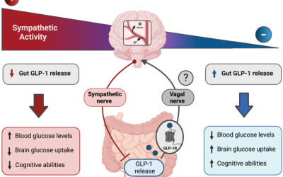
The brain-gut neurocircuitry is proving to be finely involved in a wide range of physiological functions. In this issue of Neuron, Ren et al.(1) show that adrenergic signaling suppresses postprandial glucagon-like peptide 1 (GLP-1) secretion. This, in turn, raises circulating glucose levels and impairs brain glucose uptake and cognitive function.


05/03/2024 | J Neurol Neurosurg Psychiatry
Resective epilepsy surgery and its impact on depression in adults: a systematic review, meta-analysis, and implications for future research.
Hernandez Poblete N, Gay F, Salvo F, Micoulaud-Franchi JA, Bienvenu T, Coelho J, Aupy J
doi: 10.1136/jnnp-2023-333073

Abstract:
BACKGROUND: How epilepsy surgery influences the bidirectional relationship of epilepsy and depression remains poorly defined. METHOD: For a better understanding of this question, we conducted a systematic review and meta-analysis of risk ratio on depression prevalence before and after epilepsy surgery, using Preferred Reporting Items for Systematic reviews and Meta-Analyses guidelines. Three databases were comprehensively screened for all studies assessing depression before and after resective surgery in adult epileptic patients until 8 October 2022. Studies were included if depression was assessed before and after epilepsy surgery regardless of the time of follow-up. A total of 1917 studies were screened for eligibility and 91 full-texts up for inclusion; 35 studies were finally included, 25 studies and 2563 patients were included in main meta-analysis and 10 for exploratory analysis. Risk of bias was assessed using Risk Of Bias In Non-randomised Studies - of Interventions (ROBINS-I) from Cochrane. To derive the pooled depression rates before and after surgery, a meta-analysis with inversed-variance was performed using random-effects logistic models with Peto's correction and a 95% CI. Heterogeneity was assessed with Cochran's Q-test along with its derived measure of inconsistency I(2). RESULTS: Overall, the depression rates before and after resective epilepsy surgery were 0.70 (0.53 to 0.91) 95% CI, suggesting that the rate of depression at last follow-up evaluation tends to decrease after Resective Epilepsy Surgery (RES). Subgroup analysis suggest a positive long-term effect appears with a significant lower rates of depression already 6 months (0.61 (0.38 to 0.98)), after surgery which is maintained over time after 1 year (0.53 (0.31 to 0.90)), and after 2 years (0.62 (0.42 to 0.92)). CONCLUSION: This important finding should be taken in consideration before resective surgery for drug-resistant epilepsies. However, prospective studies should be conducted to characterise which patient, at the individual level, might be at risk of de novo or worsening of depression. PROSPERO REGISTRATION NUMBER: CRD42022355386.


29/02/2024 | Sci Rep
Genetic labeling of embryonically-born dentate granule neurons in young mice using the Penk(Cre) mouse line.
Mortessagne P, Cartier E, Balia M, Fevre M, Corailler F, Herry C, Abrous DN, Battefeld A, Pacary E
doi: 10.1038/s41598-024-55299-9

Abstract:
The dentate gyrus (DG) of the hippocampus is a mosaic of dentate granule neurons (DGNs) accumulated throughout life. While many studies focused on the morpho-functional properties of adult-born DGNs, much less is known about DGNs generated during development, and in particular those born during embryogenesis. One of the main reasons for this gap is the lack of methods available to specifically label and manipulate embryonically-born DGNs. Here, we have assessed the relevance of the Penk(Cre) mouse line as a genetic model to target this embryonically-born population. In young animals, Penk(Cre) expression allows to tag neurons in the DG with positional, morphological and electrophysiological properties characteristic of DGNs born during the embryonic period. In addition, Penk(Cre)+ cells in the DG are distributed in both blades along the entire septo-temporal axis. This model thus offers new possibilities to explore the functions of this underexplored population of embryonically-born DGNs.


09/02/2024 | Transl Psychiatry
Individual variations in motives for nicotine self-administration in male rats: evidence in support for a precision psychopharmacology.
Garcia-Rivas V, Fiancette JF, Tostain J, de Maio G, Ceau M, Wiart JF, Gaulier JM, Deroche-Gamonet V
doi: 10.1038/s41398-024-02774-6

Abstract:
The significant heterogeneity in smoking behavior among smokers, coupled with the inconsistent efficacy of approved smoking cessation therapies, supports the presence of individual variations in the mechanisms underlying smoking. This emphasizes the need to shift from standardized to personalized smoking cessation therapies. However, informed precision medicine demands precision fundamental research. Tobacco smoking is influenced and sustained by diverse psychopharmacological interactions between nicotine and environmental stimuli. In the classical experimental rodent model for studying tobacco dependence, namely intravenous self-administration of nicotine, seeking behavior is reinforced by the combined delivery of nicotine and a discrete cue (nicotine+cue). Whether self-administration behavior is driven by the same psychopharmacological mechanisms across individual rats remains unknown and unexplored. To address this, we employed behavioral pharmacology and unbiased cluster analysis to investigate individual differences in the mechanisms supporting classical intravenous nicotine self-administration (0.04 mg/kg/infusion) in male outbred Sprague-Dawley rats. Our analysis identified two clusters: one subset of rats sought nicotine primarily for its reinforcing effects, while the second subset sought nicotine to enhance the reinforcing effects of the discrete cue. Varenicline (1 mg/kg i.p.) reduced seeking behavior in the former group, whereas it tended to increase in the latter group. Crucially, despite this fundamental qualitative difference revealed by behavioral manipulation, the two clusters exhibited quantitatively identical nicotine+cue self-administration behavior. The traditional application of rodent models to study the reinforcing and addictive effects of nicotine may mask individual variability in the underlying motivational mechanisms. Accounting for this variability could significantly enhance the predictive validity of translational research.


09/02/2024 | Nat Commun
Endogenous cannabinoids in the piriform cortex tune olfactory perception.
Terral G, Harrell E, Lepousez G, Wards Y, Huang D, Dolique T, Casali G, Nissant A, Lledo PM, Ferreira G, Marsicano G, Roux L
doi: 10.1038/s41467-024-45161-x

Abstract:
Sensory perception depends on interactions between external inputs transduced by peripheral sensory organs and internal network dynamics generated by central neuronal circuits. In the sensory cortex, desynchronized network states associate with high signal-to-noise ratio stimulus-evoked responses and heightened perception. Cannabinoid-type-1-receptors (CB1Rs) - which influence network coordination in the hippocampus - are present in anterior piriform cortex (aPC), a sensory paleocortex supporting olfactory perception. Yet, how CB1Rs shape aPC network activity and affect odor perception is unknown. Using pharmacological manipulations coupled with multi-electrode recordings or fiber photometry in the aPC of freely moving male mice, we show that systemic CB1R blockade as well as local drug infusion increases the amplitude of gamma oscillations in aPC, while simultaneously reducing the occurrence of synchronized population events involving aPC excitatory neurons. In animals exposed to odor sources, blockade of CB1Rs reduces correlation among aPC excitatory units and lowers behavioral olfactory detection thresholds. These results suggest that endogenous endocannabinoid signaling promotes synchronized population events and dampen gamma oscillations in the aPC which results in a reduced sensitivity to external sensory inputs.


Abstract:
BACKGROUND: Delayed cerebral ischemia associated with cerebral vasospasm (CVS) in aneurysmal subarachnoid hemorrhage significantly affects patient prognosis. Levosimendan has emerged as a potential treatment, but clinical data are lacking. The aim of this study is to decipher levosimendan's effect on cerebral hemodynamics by automated quantitative measurements of brain computed tomography perfusion (CTP). METHODS: We conducted a retrospective analysis of a database of a neurosurgical intensive care unit. All patients admitted from January 2018 to July 2022 for aneurysmal subarachnoid hemorrhage and treated with levosimendan for CVS who did not respond to other therapies were included. Quantitative measurements of time to maximum (Tmax), relative cerebral blood volume (rCBV), and relative cerebral blood flow (rCBF) were automatically compared with coregistered CTP before and after levosimendan administration in oligemic regions. RESULTS: Of 21 patients included, CTP analysis could be performed in 16. Levosimendan improved Tmax from 14.4 s (interquartile range [IQR] 9.1-21) before treatment to 7.1 s (IQR 5.5-8.1) after treatment (p < 0.001). rCBV (94% [IQR 79-103] before treatment and 89% [IQR 72-103] after treatment, p = 0.63) and rCBF (85% [IQR 77-90] before treatment and 87% [IQR 73-98] after treatment, p = 0.98) remained stable. The subgroup of six patients who did not develop cerebral infarction attributed to delayed cerebral ischemia showed an approximately 10% increase (rCBV 85% [IQR 79-99] before treatment vs. 95% [IQR 88-112] after treatment, p = 0.21; rCBF 81% [IQR 76-87] before treatment vs. 89% [IQR 84-99] after treatment, p = 0.4). CONCLUSIONS: In refractory CVS, levosimendan use was associated with a significant reduction in Tmax in oligemic regions. However, this value remained at an abnormal level, indicating the presence of a persistent CVS. Further analysis raised the hypothesis that levosimendan causes cerebral vasodilation, but other studies are needed because our design does not allow us to quantify the effect of levosimendan from that of the natural evolution of CVS.


02/02/2024 | ann intensive care
Critically ill patients with infective endocarditis, neurological complications and indication for cardiac surgery: a multicenter propensity-adjusted study.
Gros A, Seguy B, Bonnet G, Guettard YO, Pillois X, Prevel R, Orieux A, Ternacle J, Preau S, Lavie-Badie Y, Coupez E, Coudroy R, Marest D, Martins RP, Gruson D, Tourdias T, Boyer A

Abstract:
BACKGROUND: The benefit-risk balance and optimal timing of surgery for severe infective endocarditis (IE) with ischemic or hemorrhagic strokes is unknown. The study aim was to compare the neurological outcome between patients receiving surgery or not. METHODS: In a prospective register-based multicenter ICU study, patients were included if they met the following criteria: (i) left-sided IE with an indication for heart surgery; (ii) with cerebral complications documented by cerebral imaging before cardiac surgery; (iii) with Sequential Organ Failure Assessment score >/= 3. Exclusion criteria were isolated right-sided IE, in-hospital acquired IE and patients with cerebral complications only after cardiac surgery. In the primary analysis, the prognostic value of surgery in term of disability at 6 month was assessed by using a propensity score-adjusted logistic regression. RESULTS: 192 patients were included including ischemic stroke (74.5%) and hemorrhagic lesion (15.6%): 67 (35%) had medical treatment and 125 (65%) cardiac surgery. In the propensity score-adjusted logistic regression, a favorable 6-month neurological outcome was associated with surgery (odds ratio 13.8 (95% CI 6.2-33.7). The 1-year mortality was strongly reduced with surgery in the fixed-effect propensity-adjusted Cox model (hazard ratio 0.18; 95% CI 0.11-0.27; p < 0.001). These effects remained whether the patients received delayed surgery (n = 62/125) or not and whether they were deeply comatose (Glasgow Coma Scale </= 10) or not. CONCLUSIONS: In critically ill IE patients with an indication for surgery and previous cerebral events, a better propensity-adjusted neurological outcome was associated with surgery compared with medical treatment.


09/01/2024 | genomics
Cell specification and functional interactions in the pig blastocyst inferred from single-cell transcriptomics and uterine fluids proteomics.
Dufour A, Kurylo C, Stöckl JB, Laloë D, Bailly Y, Manceau P, Martins F, Turhan AG, Ferchaud S, Pain B, Fröhlich T, Foissac S, Artus J, Acloque H

Abstract:
The embryonic development of the pig comprises a long in utero pre- and peri-implantation development, which dramatically differs from mice and humans. During this peri-implantation period, a complex series of paracrine signals establishes an intimate dialogue between the embryo and the uterus. To better understand the biology of the pig blastocyst during this period, we generated a large dataset of single-cell RNAseq from early and hatched blastocysts, spheroid and ovoid conceptus and proteomic datasets from corresponding uterine fluids. Our results confirm the molecular specificity and functionality of the three main cell populations. We also discovered two previously unknown subpopulations of the trophectoderm, one characterised by the expression of LRP2, which could represent progenitor cells, and the other, expressing pro-apoptotic markers, which could correspond to the Rauber's layer. Our work provides new insights into the biology of these populations, their reciprocal functional interactions, and the molecular dialogue with the maternal uterine environment.


09/01/2024 | J Clin Endocrinol Metab
Quantitative MRI biomarkers measure changes in targeted brain areas in patients with obesity.
Baynat L, Yamamoto T, Tourdias T, Zhang B, Prevost V, Infante A, Klein A, Caid J, Cadart O, Dousset V, Gatta Cherifi B
doi: 10.1210/clinem/dgae014

Abstract:
CONTEXT: Obesity is accompanied by damages to several tissues, including the brain. Pathological data and animal models have demonstrated an increased inflammatory reaction in hypothalamus and hippocampus. OBJECTIVE: We tested whether we could observe such pathological modifications in vivo through quantitative MRI metrics. DESIGN: This prospective study was conducted between May 2019 and November 2022. SETTING: The study was conducted in the Specialised Center for the Care of Obesity in a French Universitary Hospital. PATIENTS: Twenty seven patients with obesity and 23 age and gender-paired normal-weight controls were prospectively recruited. INTERVENTIONS: All participants were explored on brain MRI. Anthropometric and biological data, eating behavior, anxiety, depression and memory performance were assessed on both groups. MAIN OUTCOME MEASURE: The main outcome measure was brain MRI with the following parametric maps: quantitative susceptibility mapping (QSM), mean diffusivity (MD), fractional anisotropy (FA), magnetization transfer ratio map (MTR) and T2 relaxivity map (R2). RESULTS: In the hypothalamus, patients with obesity had higher FA, lower QSM compared to normal-weight controls. In the hippocampus, patients with obesity had higher FA and lower MD. There was no correlation between imaging biomarkers and eating behavior or anxiety. CONCLUSION: Our findings are consistent with the presence of neuro-inflammation in brain regions involved in food intake. In vivo brain biomarkers from quantitative MRI appear to provide an incremental information for the assessment of brain damages in patients with obesity.


26/12/2023 | biomedicines
COVID-19 on Oral Health: A New Bilateral Connection for the Pandemic.
Bellocchio L, Dipalma G, Inchingolo AM, Inchingolo AD, Ferrante L, Del Vecchio G, Malcangi G, Palermo A, Qendro A, Inchingolo F
doi: 10.3390/biomedicines12010060

Abstract:
Severe acute respiratory syndrome coronavirus 2 (SARS-CoV-2) infection and transmission are generally known to be produced by respiratory droplets and aerosols from the oral cavity (O.C.) of infected subjects, as stated by the World Health Organization. Saliva also retains the viral particles and aids in the spread of COVID-19. Angiotensin-converting enzyme Type 2 (ACE2) and transmembrane serine protease 2 (TMPRSS2) are two of the numerous factors that promote SARS-CoV-2 infection, expressed by O.C. structures, various mucosa types, and the epithelia of salivary glands. A systemic SARS-CoV-2 infection might result from viral replication in O.C. cells. On the other hand, cellular damage of different subtypes in the O.C. might be associated with various clinical signs and symptoms. Factors interfering with SARS-CoV-2 infection potential might represent fertile ground for possible local pharmacotherapeutic interventions, which may confine SARS-CoV-2 virus entry and transmission in the O.C., finally representing a way to reduce COVID-19 incidence and severity.


14/12/2023 | Nat Commun
CA3 hippocampal synaptic plasticity supports ripple physiology during memory consolidation.
El Oussini H, Zhang CL, Francois U, Castelli C, Lampin-Saint-Amaux A, Lepleux M, Molle P, Velez L, Dejean C, Lanore F, Herry C, Choquet D, Humeau Y
doi: 10.1038/s41467-023-42969-x

Abstract:
The consolidation of recent memories depends on memory replays, also called ripples, generated within the hippocampus during slow-wave sleep, and whose inactivation leads to memory impairment. For now, the mobilisation, localisation and importance of synaptic plasticity events associated to ripples are largely unknown. To tackle this question, we used cell surface AMPAR immobilisation to block post-synaptic LTP within the hippocampal region of male mice during a spatial memory task, and show that: 1- hippocampal synaptic plasticity is engaged during consolidation, but is dispensable during encoding or retrieval. 2- Plasticity blockade during sleep results in apparent forgetting of the encoded rule. 3- In vivo ripple recordings show a strong effect of AMPAR immobilisation when a rule has been recently encoded. 4- In situ investigation suggests that plasticity at CA3-CA3 recurrent synapses supports ripple generation. We thus propose that post-synaptic AMPAR mobility at CA3 recurrent synapses is necessary for ripple-dependent rule consolidation.


01/12/2023 | Hum Brain Mapp
Lifespan neurodegeneration of the human brain in multiple sclerosis.
Coupe P, Planche V, Mansencal B, Kamroui RA, Koubiyr I, Manjon JV, Tourdias T

Abstract:
Atrophy related to multiple sclerosis (MS) has been found at the early stages of the disease. However, the archetype dynamic trajectories of the neurodegenerative process, even prior to clinical diagnosis, remain unknown. We modeled the volumetric trajectories of brain structures across the entire lifespan using 40,944 subjects (38,295 healthy controls and 2649 MS patients). Then, we estimated the chronological progression of MS by assessing the divergence of lifespan trajectories between normal brain charts and MS brain charts. Chronologically, the first affected structure was the thalamus, then the putamen and the pallidum (around 4 years later), followed by the ventral diencephalon (around 7 years after thalamus) and finally the brainstem (around 9 years after thalamus). To a lesser extent, the anterior cingulate gyrus, insular cortex, occipital pole, caudate and hippocampus were impacted. Finally, the precuneus and accumbens nuclei exhibited a limited atrophy pattern. Subcortical atrophy was more pronounced than cortical atrophy. The thalamus was the most impacted structure with a very early divergence in life. Our experiments showed that lifespan models of most impacted structures could be an important tool for future preclinical/prodromal prognosis and monitoring of MS.


12/2023 | Nat Med
Thalamic deep brain stimulation in traumatic brain injury: a phase 1, randomized feasibility study.
Schiff ND, Giacino JT, Butson CR, Choi EY, Baker JL, O'Sullivan KP, Janson AP, Bergin M, Bronte-Stewart HM, Chua J, DeGeorge L, Dikmen S, Fogarty A, Gerber LM, Krel M, Maldonado J, Radovan M, Shah SA, Su J, Temkin N, Tourdias T, Victor JD, Waters A, Kolakowsky-Hayner SA, Fins JJ, Machado AG, Rutt BK, Henderson JM
doi: 10.1038/s41591-023-02638-4

Abstract:
Converging evidence indicates that impairments in executive function and information-processing speed limit quality of life and social reentry after moderate-to-severe traumatic brain injury (msTBI). These deficits reflect dysfunction of frontostriatal networks for which the central lateral (CL) nucleus of the thalamus is a critical node. The primary objective of this feasibility study was to test the safety and efficacy of deep brain stimulation within the CL and the associated medial dorsal tegmental (CL/DTTm) tract.Six participants with msTBI, who were between 3 and 18 years post-injury, underwent surgery with electrode placement guided by imaging and subject-specific biophysical modeling to predict activation of the CL/DTTm tract. The primary efficacy measure was improvement in executive control indexed by processing speed on part B of the trail-making test.All six participants were safely implanted. Five participants completed the study and one was withdrawn for protocol non-compliance. Processing speed on part B of the trail-making test improved 15% to 52% from baseline, exceeding the 10% benchmark for improvement in all five cases.CL/DTTm deep brain stimulation can be safely applied and may improve executive control in patients with msTBI who are in the chronic phase of recovery.ClinicalTrials.gov identifier: NCT02881151 .


Abstract:
Excessive neural variability of sensory responses is a hallmark of atypical sensory processing in autistic individuals with cascading effects on other core autism symptoms but unknown neurobiological substrate. Here, by recording neocortical single neuron activity in a well-established mouse model of Fragile X syndrome and autism, we characterized atypical sensory processing and probed the role of endogenous noise sources in exaggerated response variability in males. The analysis of sensory stimulus evoked activity and spontaneous dynamics, as well as neuronal features, reveals a complex cellular and network phenotype. Neocortical sensory information processing is more variable and temporally imprecise. Increased trial-by-trial and inter-neuronal response variability is strongly related to key endogenous noise features, and may give rise to behavioural sensory responsiveness variability in autism. We provide a novel preclinical framework for understanding the sources of endogenous noise and its contribution to core autism symptoms, and for testing the functional consequences for mechanism-based manipulation of noise.


04/11/2023 | J Endocrinol Invest
Endocannabinoid-related molecules predict the metabolic efficacy of GLP-1 receptor agonism in humans with obesity.
Matias I, Lehmann EW, Zizzari P, Byberg S, Cota D, Torekov SS, Quarta C
doi: 10.1007/s40618-023-02228-8

Abstract:
OBJECTIVE: N-acylethanolamines (NAEs) include endocannabinoid (EC) and EC-related molecules that impact the anti-obesity and anti-diabetic efficacy of glucagon-like peptide-1 receptor agonists (GLP-1RA) in animal studies. However, the clinical relevance of these findings remains to be determined. Here, we tested whether GLP-1RA treatment affects circulating NAE levels and whether NAEs may predict the efficacy of GLP-1RA treatment in humans with obesity undergoing weight loss maintenance. MATERIALS AND METHODS: We profiled plasma levels of NAEs in participants with obesity undergoing weight loss maintenance with (n = 23)/or without (n = 20) treatment with the GLP-1RA liraglutide. NAE levels were measured at three different time points: before the start of the study, at the end of the diet-induced weight loss, and after 52-weeks treatment. Linear regression analyses were used to investigate whether pharmacological responses could be predicted by NAEs levels. RESULTS: Liraglutide treatment reduced plasma concentrations of the NAE and oleoyl-ethanolamide (OEA), without altering arachidonoyl-ethanolamide (AEA) levels and palmitoyl-ethanolamide (PEA) levels. High pre-treatment levels of OEA were predictive of superior compound-mediated effects on fasting insulin and triglyceride levels. High pre-treatment PEA and AEA levels were also predictive of superior Liraglutide-mediated effects on triglyceride levels. CONCLUSIONS: Our data suggests that specific NAEs such as OEA and AEA are promising biomarkers of GLP-1RA metabolic efficacy in humans with obesity during weight loss maintenance. Plasma profiling of EC-related molecules may be a promising strategy to tailor GLP-1R-based therapies to individual needs in obesity and diabetes management.


11/2023 | j neurointerv surg
Influence of prior intravenous thrombolysis in patients treated with mechanical thrombectomy for M2 occlusions: insight from the Endovascular Treatment in Ischemic Stroke (ETIS) registry.
Le Floch A, Clarencon F, Rouchaud A, Kyheng M, Labreuche J, Sibon I, Boulouis G, Gory B, Richard S, Caroff J, Blanc R, Seners P, Eker OF, Cho TH, Consoli A, Bourcier R, Guillon B, Dargazanli C, Arquizan C, Denier C, Eugene F, Vannier S, Gentric JC, Gauberti M, Naggara O, Rosso C, Turc G, Ozkul-Wermester O, Cognard C, Albucher JF, Timsit S, Bourdain F, Le Bras A, Richter S, Moulin S, Pop R, Heck O, Moreno R, L'Allinec V, Lapergue B, Marnat G
doi: 10.1136/jnis-2022-019672

Abstract:
BACKGROUND: Intravenous thrombolysis (IVT) for patients treated with mechanical thrombectomy (MT) for proximal occlusions has recently been questioned through randomized trials. However, few patients with M2 occlusions were included. We investigated the influence of prior IVT for patients presenting M2 occlusions treated with MT in comparison with MT alone. METHODS: We conducted a retrospective analysis of the Endovascular Treatment in Ischemic Stroke (ETIS) registry, a multicenter observational study. Data from consecutive patients treated with MT for M2 occlusions between January 2015 and January 2022 at 26 comprehensive stroke centers were analyzed. The primary endpoint was 90-day modified Rankin Scale score of 0-2. Outcomes were compared using propensity score approaches. We also performed sensitivity analysis in relevant subgroups of patients. RESULTS: Among 1132 patients with M2 occlusions treated with MT, 570 received prior IVT. The two groups were comparable after propensity analysis. The rate of favorable functional outcome was significantly higher in the IVT+MT group compared with the MT alone group (59.8% vs 44.7%; adjusted OR 1.38, 95% CI 1.10 to 1.75, P=0.008). Hemorrhagic and procedural complications were similar in both groups. In sensitivity analysis excluding patients with anticoagulation treatment, favorable recanalization was more frequent in the IVT+MT group (OR 1.37, 95% CI 1.11 to 1.70, P=0.004). CONCLUSIONS: In cases of M2 occlusions, prior IVT combined with MT resulted in better functional outcome than MT alone, without increasing the rate of hemorrhagic or procedural complications. These results suggest the benefit of IVT in patients undergoing MT for M2 occlusions.


30/10/2023 | Nat Neurosci
Prefrontal circuits encode both general danger and specific threat representations.
Martin-Fernandez M, Menegolla AP, Lopez-Fernandez G, Winke N, Jercog D, Kim HR, Girard D, Dejean C, Herry C
doi: 10.1038/s41593-023-01472-8

Abstract:
Behavioral adaptation to potential threats requires both a global representation of danger to prepare the organism to react in a timely manner but also the identification of specific threatening situations to select the appropriate behavioral responses. The prefrontal cortex is known to control threat-related behaviors, yet it is unknown whether it encodes global defensive states and/or the identity of specific threatening encounters. Using a new behavioral paradigm that exposes mice to different threatening situations, we show that the dorsomedial prefrontal cortex (dmPFC) encodes a general representation of danger while simultaneously encoding a specific neuronal representation of each threat. Importantly, the global representation of danger persisted in error trials that instead lacked specific threat identity representations. Consistently, optogenetic prefrontal inhibition impaired overall behavioral performance and discrimination of different threatening situations without any bias toward active or passive behaviors. Together, these data indicate that the prefrontal cortex encodes both a global representation of danger and specific representations of threat identity to control the selection of defensive behaviors.


20/10/2023 | Curr Biol
Striatopallidal cannabinoid type-1 receptors mediate amphetamine-induced sensitization.
Mariani Y, Covelo A, Rodrigues RS, Julio-Kalajzic F, Pagano Zottola AC, Lavanco G, Fabrizio M, Gisquet D, Drago F, Cannich A, Baufreton J, Marsicano G, Bellocchio L
doi: 10.1016/j.cub.2023.09.075

Abstract:
Repeated exposure to psychostimulants, such as amphetamine, causes a long-lasting enhancement in the behavioral responses to the drug, called behavioral sensitization.(1) This phenomenon involves several neuronal systems and brain areas, among which the dorsal striatum plays a key role.(2) The endocannabinoid system (ECS) has been proposed to participate in this effect, but the neuronal basis of this interaction has not been investigated.(3) In the CNS, the ECS exerts its functions mainly acting through the cannabinoid type-1 (CB(1)) receptor, which is highly expressed at terminals of striatal medium spiny neurons (MSNs) belonging to both the direct and indirect pathways.(4) In this study, we show that, although striatal CB(1) receptors are not involved in the acute response to amphetamine, the behavioral sensitization and related synaptic changes require the activation of CB(1) receptors specifically located at striatopallidal MSNs (indirect pathway). These results highlight a new mechanism of psychostimulant sensitization, a phenomenon that plays a key role in the health-threatening effects of these drugs.